

Endoscopic resection of advanced ampullary adenomas: a single-center 14-year retrospective cohort study
- PMID: 30167949
- PMCID: PMC6430826
- DOI: 10.1007/s00464-018-6392-9
Endoscopic resection of advanced ampullary adenomas: a single-center 14-year retrospective cohort study
Abstract
Background: Endoscopic ampullectomy has been recognized as a safe and reliable means to resect selective tumors of the ampulla of Vater and is associated with lower morbidity and mortality rates compared to surgical resection. Success rates range from 42 to 92%, with recurrences reported in up to 33%. Studies on endoscopic resection of advanced lesions such as those with intraductal extension of adenoma (IEA) and lateral spreading adenomas (LSA) are limited. We aimed to evaluate the technical success, complications, and recurrence of endoscopic resection of ampullary adenomas, including advanced lesions.
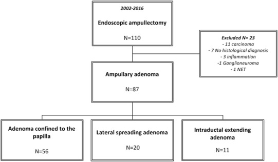
Methods: All patients referred to the Erasmus Medical Center for endoscopic resection of an ampullary lesion were retrospectively identified between 2002 and 2016. Endoscopic success was defined as complete excision of the adenoma, irrespective of the number of attempts, in the absence of recurrence.
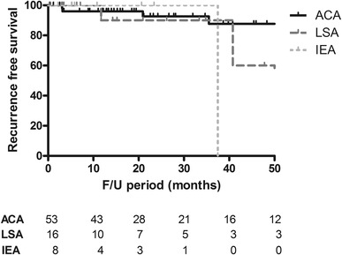
Results: We included 87 patients with a median age of 65 years. Of these, 56 patients (64%) had an adenoma confined to the ampulla (ACA), 20 patients (23%) had an LSA, and 11 patients (13%) were treated for an IEA. The median lesion sizes were 24.6 mm, 41.4 mm, and 16.3 mm, respectively (P < 0.001). Complications occurred in 22 patients (25.3%), of which hemorrhage was most prevalent (12.6%), followed by perforation (8.1%). Complications were equally divided (P = 0.874). The median follow-up duration was 21.1 months (12-45.9) for ACA, 14.7 months (4.2-34.5) for LSA, and 5.8 months (3.7-22.0) for IEA (P = 0.051). Endoscopic resection was curative in 87.5% of patients with an ACA, 85% in patients with an LSA, and in only one patient with an IEA (P < 0.001). Recurrence occurred in 10 patients (11.5%) (P = 0.733).
Conclusion: Endoscopic ampullectomy is safe and highly successful in selected patients with an adenoma with or without lateral spreading. Outcomes of endoscopic treatment adenomas with an intraductal extension are less favorable and in these cases surgery should be considered.
Keywords: Ampulla of Vater; Ampullary adenoma; ERCP; Endoscopic ampullectomy; Endoscopic resection.
Conflict of interest statement
Dr. J.W. Poley reports personal fees from Boston Scientific, personal fees from Cook Medical and personal fees from Pentax, outside the submitted work. Dr. Bruno reports grants from Boston Scientific, personal fees from Boston Scientific, grants from Cook Medical, personal fees from Cook Medical, grants from 3M and personal fees from 3M, outside the submitted work. S.E. van der Wiel and Dr. A.D. Koch have no conflicts of interest of financial ties to disclose.
Figures
Similar articles
-
Outcomes after endoscopic resection of large laterally spreading lesions of the papilla and conventional ampullary adenomas are equivalent.Endoscopy. 2018 Oct;50(10):972-983. doi: 10.1055/a-0587-5228. Epub 2018 May 16. Endoscopy. 2018. PMID: 29768645
-
Endoscopic resection of ampullary neoplasms: a single-center experience.Surg Endosc. 2009 Nov;23(11):2568-74. doi: 10.1007/s00464-009-0464-9. Epub 2009 Apr 10. Surg Endosc. 2009. PMID: 19360365
-
Endoscopic resection of ampullary lesions: a single-center 8-year retrospective cohort study of 91 patients with long-term follow-up.Surg Endosc. 2013 Oct;27(10):3865-76. doi: 10.1007/s00464-013-2996-2. Epub 2013 May 25. Surg Endosc. 2013. PMID: 23708714
-
Endoscopic ampullectomy: techniques and outcomes.J Clin Gastroenterol. 2012 Jan;46(1):8-15. doi: 10.1097/MCG.0b013e318233a844. J Clin Gastroenterol. 2012. PMID: 22064552 Review.
-
Long-term recurrence after endoscopic versus surgical ampullectomy of sporadic ampullary adenomas: a systematic review and meta-analysis.Surg Endosc. 2023 Jul;37(7):5022-5044. doi: 10.1007/s00464-023-10083-0. Epub 2023 May 23. Surg Endosc. 2023. PMID: 37221416
Cited by
-
Expert consensus on endoscopic papillectomy using a Delphi process.Gastrointest Endosc. 2021 Oct;94(4):760-773.e18. doi: 10.1016/j.gie.2021.04.009. Epub 2021 Apr 19. Gastrointest Endosc. 2021. PMID: 33887269 Free PMC article.
-
Management of Remnant or Recurrent Lesions after Endoscopic Papillectomy.Clin Endosc. 2020 Nov;53(6):659-662. doi: 10.5946/ce.2019.171. Epub 2019 Dec 3. Clin Endosc. 2020. PMID: 31794653 Free PMC article.
-
Effectiveness of endoscopic papillectomy with stent placement in pancreatic and bile ducts for treating duodenal papillary adenoma: a retrospective study.BMC Gastroenterol. 2024 Oct 24;24(1):379. doi: 10.1186/s12876-024-03466-7. BMC Gastroenterol. 2024. PMID: 39448906 Free PMC article.
-
Systematic Review with Meta-Analysis: Endoscopic and Surgical Resection for Ampullary Lesions.J Clin Med. 2020 Nov 10;9(11):3622. doi: 10.3390/jcm9113622. J Clin Med. 2020. PMID: 33182806 Free PMC article. Review.
-
Comparative analysis of differences for pathological upgrade and incomplete resection in endoscopic snare papillectomy for ampullary adenomas: A single-institution retrospective study.PLoS One. 2025 Aug 12;20(8):e0330220. doi: 10.1371/journal.pone.0330220. eCollection 2025. PLoS One. 2025. PMID: 40794694 Free PMC article.
References
MeSH terms
LinkOut - more resources
Full Text Sources
Other Literature Sources
