

Vascular wall imaging in reversible cerebral vasoconstriction syndrome - a 3-T contrast-enhanced MRI study
- PMID: 30167985
- PMCID: PMC6117223
- DOI: 10.1186/s10194-018-0906-7
Vascular wall imaging in reversible cerebral vasoconstriction syndrome - a 3-T contrast-enhanced MRI study
Abstract
Background: Limited histopathology studies have suggested that reversible cerebral vasoconstriction syndromes (RCVS) does not present with vascular wall inflammation. Previous vascular imaging studies have had inconsistent vascular wall enhancement findings in RCVS patients. The aim of this study was to determine whether absence of arterial wall pathology on imaging is a universal finding in patients with RCVS.
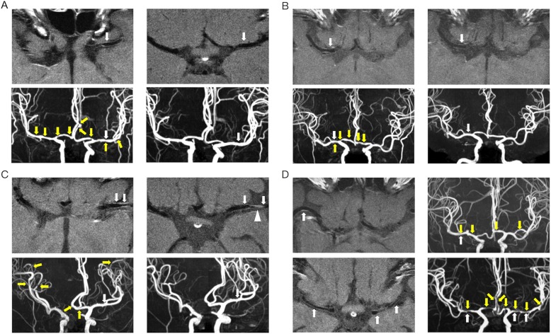
Methods: We recruited patients with RCVS from Taipei Veterans General Hospital prospectively from 2010 to 2012, with follow-up until 2017 (n = 48). We analyzed the characteristics of vascular wall enhancement in these patients without comparisons to a control group. All participants received vascular wall imaging by contrasted T1 fluid-attenuated inversion recovery with a 3-T magnetic resonance machine. The vascular wall enhancement was rated as marked, mild or absent.
Results: Of 48 patients with RCVS, 22 (45.8%) had vascular wall enhancement (5 marked and 17 mild). Demographics, clinical profiles, and cerebral artery flow velocities were similar across patients with versus without vascular wall enhancement, except that patients with vascular wall enhancement had fewer headache attacks than those without (p = 0.04). Follow-up imaging completed in 14 patients (median interval, 7 months) showed reduced enhancement in 9 patients, but persistent enhancement in 5.
Conclusion: Almost half of our RCVS patients exhibited imaging enhancement of diseased vessels, and it was persistent for approximately a third of those patients with follow-up imaging. Both acute and persistent vascular wall enhancement may be unhelpful for differentiating RCVS from central nervous system vasculitis or subclinical atherosclerosis.
Keywords: Contrast enhancement; Reversible cerebral vasoconstriction syndromes; Thunderclap headache; Vascular wall imaging.
Conflict of interest statement
Ethics approval and consent to participate
The study protocol was approved by the Taipei Veterans General Hospital Institutional Review Board. All participants provided written informed consent before entering the study. All clinical investigations were conducted according to the principles of the Declaration of Helsinki. The corresponding author had full access to all the data in the study and had final responsibility for the decision to submit for publication.
Consent for publication
Not applicable
Competing interests
The authors declare that they have no competing interests.
Publisher’s Note
Springer Nature remains neutral with regard to jurisdictional claims in published maps and institutional affiliations.
Figures
References
MeSH terms
Substances
Grants and funding
- The Featured Areas Research Center Program within the framework of the Higher Education Sprout Project by the Ministry of Education (MOE)/Brain Research Center, National Yang-Ming University
- V100E6-001, VGHUST105-G7-1-1, V105C-127, V105E9-001-MY2-1, VTA105-V1-1-1/Taipei Veterans General Hospital
- V106C-117/Taipei Veterans General Hospital
- MOST 106-2314-B-010 -019 -MY2, MOST 107-2321-B-010 -001 -, and MOST 104-2314-B-010 -071 -MY3/Ministry of Science and Technology, Taiwan
- IBMS-CRC103-P04/Academia Sinica
LinkOut - more resources
Full Text Sources
Other Literature Sources
Medical
