. 2018 Dec;17(6):e12833.
doi: 10.1111/acel.12833. Epub 2018 Aug 31.

Inflammaging impairs peripheral nerve maintenance and regeneration

Affiliations

Inflammaging impairs peripheral nerve maintenance and regeneration

Robert Büttner et al. Aging Cell. 2018 Dec.

Abstract

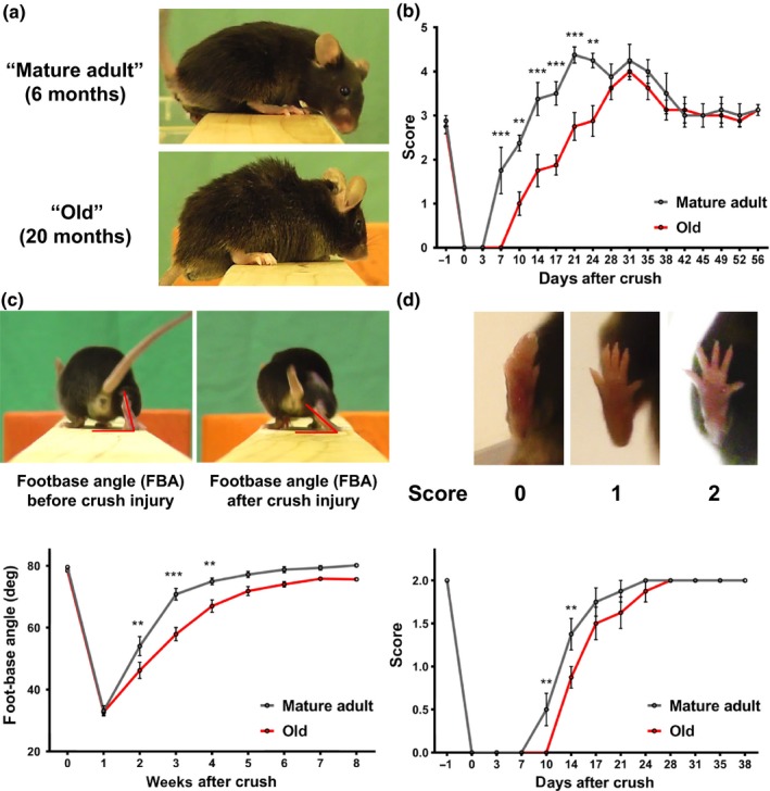
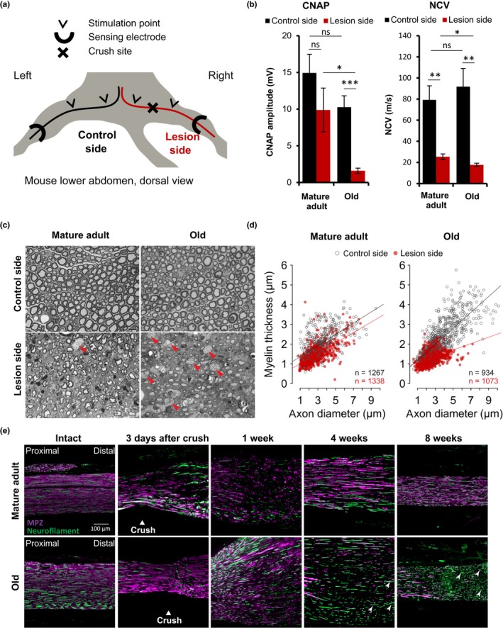
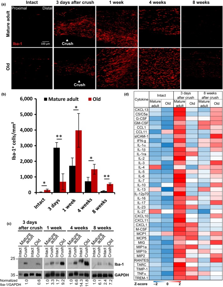
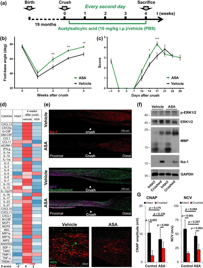
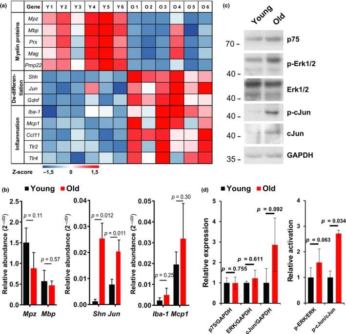
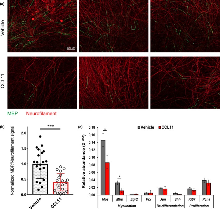
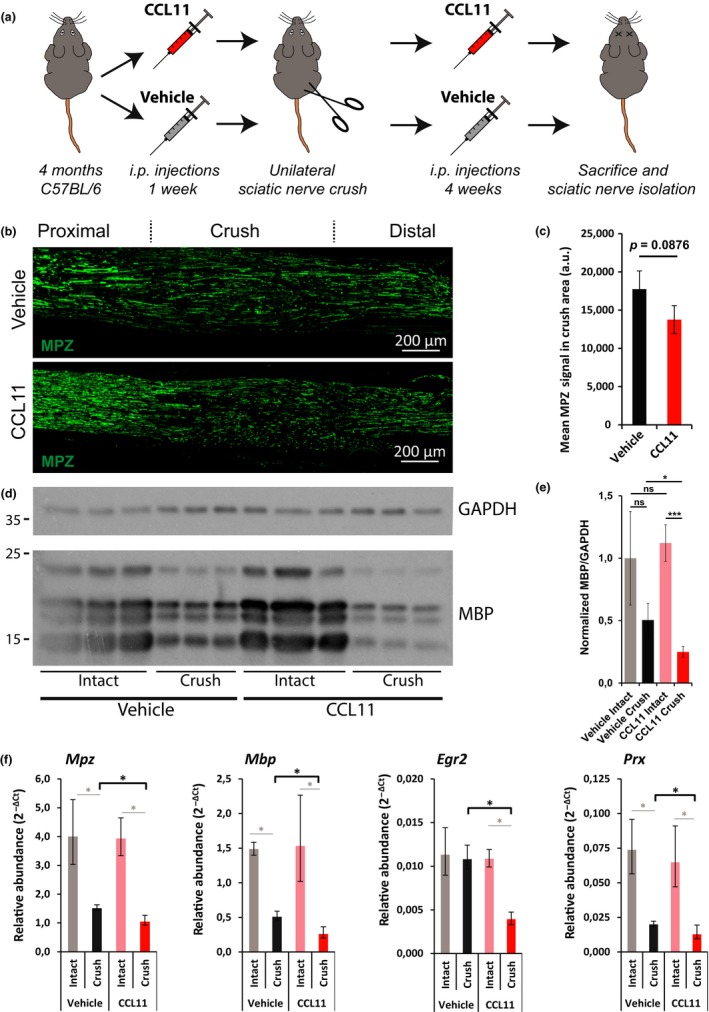
The regenerative capacity of peripheral nerves declines during aging, contributing to the development of neuropathies, limiting organism function. Changes in Schwann cells prompt failures in instructing maintenance and regeneration of aging nerves; molecular mechanisms of which have yet to be delineated. Here, we identified an altered inflammatory environment leading to a defective Schwann cell response, as an underlying mechanism of impaired nerve regeneration during aging. Chronic inflammation was detected in intact uninjured old nerves, characterized by increased macrophage infiltration and raised levels of monocyte chemoattractant protein 1 (MCP1) and CC chemokine ligand 11 (CCL11). Schwann cells in the old nerves appeared partially dedifferentiated, accompanied by an activated repair program independent of injury. Upon sciatic nerve injury, an initial delayed immune response was followed by a persistent hyperinflammatory state accompanied by a diminished repair process. As a contributing factor to nerve aging, we showed that CCL11 interfered with Schwann cell differentiation in vitro and in vivo. Our results indicate that increased infiltration of macrophages and inflammatory signals diminish regenerative capacity of aging nerves by altering Schwann cell behavior. The study identifies CCL11 as a promising target for anti-inflammatory therapies aiming to improve nerve regeneration in old age.

Keywords: aging; inflammaging; macrophages; neural regeneration; peripheral nervous system; schwann cell.


Figures

Figure 1
Figure 1
Aging impairs functional recovery after sciatic nerve crush injury. (a) “Mature adult” (6 months) and “old” (20 months) mice were subjected to sciatic nerve crush injury, and regeneration was assessed by monitoring recovery of sensory and motor functions. (b) Sensory recovery was tested by responsiveness of the paw to monofilaments of varying stiffness. Scoring reflects bending threshold forces: (0) no response for 300 g, (1) 300 g, (2) 4 g, (3) 2 g, (4) 0.4 g, and (5) 0.07 g. Motor recovery was tested by (c) measurement of the footbase angle and (d) toe spreading. Significances of all differences were calculated by two‐way ANOVA with Holm–Sidăk post hoc test and indicated by *p < 0.05, **p < 0.01, ***p < 0.001, mean ± SEM. n = 8 mice per age in b and d, n = 7 in c
Figure 2
Figure 2
Aging impairs structural regeneration after sciatic nerve crush injury. (a) For electrophysiological measurements, mice were anesthetized and fixed in an illustrated set‐up 4 weeks after unilateral sciatic nerve crush injury. (b) Compound nerve action potentials (CNAP) and nerve conduction velocities (NCV) were measured in situ on crushed sciatic nerves and intact contralateral control nerves of six mature adult mice and eight old mice; mean ± SEM. *p < 0.05, ** p < 0.01, *** p < 0.001 by unpaired, two‐tailed t‐test. (c) Representative Toluidine blue‐stained semi‐thin cross‐sections of mature adult and old sciatic nerves 4 weeks after crush injury and intact contralateral, respectively. Cross‐sections in the crush area particularly of old sciatic nerves show multiple macrophages (red arrowheads); scale bar: 10 µm. (d) Myelin thickness relative to axon diameter was quantified in cross‐sections of uninjured (control side) and injured (lesion side) sciatic nerves of four mature adult and five old mice four weeks after crush injury and illustrated as scatter plot. Per nerve 156 to 448 axons plus myelin sheath were measured and the sum of quantified axons per age and side indicated within the plots. Linear mixed models with Tukey post hoc test indicate highly significant differences (p < 0.0001) between injured and uninjured nerves for both ages as well as between injured nerves of mature adult and old mice. (e) Timeline of representative longitudinal sciatic nerve sections at the crush site, before and at indicated time points after crush injury, immunostained for myelin protein zero (MPZ purple) to mark myelination and neurofilament (green) to mark axonal fibers; proximal left and distal right, white arrowheads point at unmyelinated axons in old mice far from the remyelination frontier, scale bar: 100 μm
Figure 3
Figure 3
Age‐related changes of the nerve injury‐induced immune response. (a) Timeline of representative longitudinal sciatic nerve sections, before and at indicated time points after crush injury, immunostained for Iba‐1 to mark macrophages; scale bar: 100 μm. (b) Quantification of Iba‐1‐positive cells per area in immunostainings of n = 3 biological replicates; mean ±SD. * p < 0.05, ** p < 0.01 by unpaired, two‐tailed t‐test. (c) Immunoblots for Iba‐1 mark macrophage presence in lysates of intact or crushed sciatic nerves at indicated time points. Lysates were pooled from n = 3 different mice for all time points. The blot of 3 days after crush is from a different gel than the other samples. Equal loading is indicated by GAPDH. (d) Heatmap with row‐specific Z‐scores of a dot‐blot array (Supporting information Figure S2A) shows cytokine expression in intact and crushed sciatic nerve lysates. Pooled lysates from n = 3 mice per age and time point.
Figure 4
Figure 4
Acetylsalicylic acid improves peripheral nerve repair in old mice. (a) Two cohorts of n = 6 mice were subjected to unilateral sciatic nerve crush injury procedure and drug therapy with acetylsalicylic acid (ASA) or PBS (vehicle). ASA (10 mg per kg body weight) or PBS was injected intraperitoneally for four weeks, beginning on day 3 after crush injury and every second day thereafter. (b, c) Recovery of motor and sensory function was assessed using single‐frame motion analysis and Semmes–Weinstein monofilament test; n = 6 mice per group (n = 5 ASA‐treated mice in SFMA), significant differences determined in two‐way ANOVA with Holm–Sidăk post hoc test, *p < 0.05, **p < 0.01, mean ± SEM. (d) Heatmap with row‐specific Z‐scores of a dot‐blot array (Supporting information Figure S2C) to measure cytokine expression in pooled sciatic nerve lysates (n = 3 mice) of mature adult and old mice, treated with ASA or vehicle control. (e) Representative immunostainings of longitudinal sciatic nerve sections at the crush area of vehicle‐ and ASA‐treated mice 4 weeks after crush injury. Immunolabeling of Iba‐1, MPZ, and neurofilament indicate macrophage appearance, myelination, and axonal fibers; proximal left and distal right, scale bar: 500 μm. Immunostainings for Arginase1 and iNOS indicate M2 and M1 macrophage populations, proximal left and distal right, scale bar: 100 µm. (F) Immunoblot analysis of Erk1/2 expression and phosphorylation, MBP (myelinating Schwann cells) and Iba‐1 (macrophages). GAPDH indicates equal loading. Pooled sciatic nerve lysates from n = 3 mice. (G) Compound nerve action potentials (CNAP) and nerve conduction velocities (NCV) were measured in situ on crushed and intact sciatic nerves. n = 6 old mice treated with ASA or vehicle for 4 weeks after crush; mean ± SD. p‐values were calculated by two‐way ANOVA with Holm–Sidăk post hoc test and indicated in the diagrams
Figure 5
Figure 5
Inflammaging in sciatic nerves of old mice correlates with insufficient repair. (a) Expression of selected genes involved in myelination, dedifferentiation, and inflammation quantified from intact sciatic nerves of n = 6 young (3 months) and old (20 months) mice by RNA‐Seq. Obtained total read counts are illustrated in a heatmap with row‐specific Z‐scores. (b) Based on RNA‐Seq data, selected marker genes for myelination (Mpz, Mbp), dedifferentiation (Shh, Jun), and inflammation (Iba-1, Mcp1) were further validated by qPCR. n = 3 biological replicates for each gene and age except Shh young, where n = 2; mean ± SD of relative abundance. Indicated p‐values calculated by unpaired, two‐tailed t‐test. (c) Immunoblots of p75, Erk1/2, and cJun protein expression and phosphorylation, that is, activation of Erk1/2 and cJun, which are involved in Schwann cell repair program control. Equal loading indicated by GAPDH. Pooled sciatic nerve lysates from n = 3 mice. (d) Quantification of relative expression and phosphorylation, that is, activation, of Erk and cJun. n = 3 biological replicates. Significant differences between means ±SEM calculated by unpaired, two‐tailed t‐test, p‐values indicated in the diagrams
Figure 6
Figure 6
CCL11 impairs Schwann cell myelination in DRG coculture. DRG‐derived neurons and Schwann cells were cocultured for 6 days in normal growth medium plus 8 days in myelination‐promoting medium, both containing CCL11 (100 ng/ml) or vehicle (0.1% BSA) in PBS. (a) Representative pictures of CCL11‐ and vehicle‐treated cocultures after myelination, stained for myelin basic protein (MBP, green) and neurofilament protein H (neurofilament, red), scale bar: 100 µm. (b) Quantification of induced myelination per neurons in CCL11‐ and vehicle‐treated cocultures by normalized ratio between MBP and neurofilament signal. n = 10 biological replicates (DRG explants) quantified per experiment in two independent experiments. Scatter plot diagram of mean ± SD of normalized ratios between MBP and neurofilament signal. ***p < 0.001 by unpaired, two‐tailed t‐test. (c) Representative quantitative qPCR for two independent experiments measuring myelination, dedifferentiation, and proliferation markers in CCL11‐ and vehicle‐treated cocultures following incubation in myelination‐promoting medium. n = 4 biological replicates; mean ± SD. *p < 0.05 by unpaired, two‐tailed t‐test
Figure 7
Figure 7
Decreased sciatic nerve remyelination in CCL11‐treated mice. (a) Scheme of in vivo experiment. One week before and four weeks after unilateral sciatic nerve crush injury, CCL11 (10 µg/kg body weight in PBS) or vehicle (PBS) was injected intraperitoneally every third to fourth day. Four weeks after crush injury, mice were sacrificed and sciatic nerves isolated. (b) Representative longitudinal sciatic nerve sections of vehicle‐ and CCL11‐treated mice four weeks after crush injury stained for myelin protein zero (MPZ, green) as marker for remyelination; crush area centered, proximal left, distal right, scale bar: 200 µm. (c) Quantification of mean MPZ signal in the crush area. n = 3 biological replicates per cohort; mean ± SD. p‐value calculated by unpaired, two‐tailed t‐test. (d) Immunoblots of MBP and GAPDH in crushed and intact sciatic nerves of n = 3 vehicle‐ and CCL11‐treated mice four weeks after injury. (e) Quantification of D. n = 3 biological replicates; mean ± SD. *p < 0.05, ***p < 0.001 with unpaired, two‐tailed t‐test. (f) Quantification of myelin protein genes expression by qPCR. n = 3 biological replicates; mean ± SD. *p < 0.05 with unpaired, two‐tailed t‐test

Similar articles

Cited by

References

    1. Arango Duque, G. , & Descoteaux, A. (2014). Macrophage cytokines: Involvement in immunity and infectious diseases. Frontiers in Immunology, 5, 491. - PMC - PubMed
    1. Arthur‐Farraj, P. J. , Latouche, M. , Wilton, D. K. , Quintes, S. , Chabrol, E. , Banerjee, A. , … Jessen, K. R. (2012). c‐ Jun reprograms Schwann cells of injured nerves to generate a repair cell essential for regeneration. Neuron, 75, 633–647. 10.1016/j.neuron.2012.06.021 - DOI - PMC - PubMed
    1. Boivin, A. , Pineau, I. , Barrette, B. , Filali, M. , Vallieres, N. , Rivest, S. , & Lacroix, S. (2007). Toll‐like receptor signaling is critical for Wallerian degeneration and functional recovery after peripheral nerve injury. The Journal of Neuroscience : the Official Journal of the Society for Neuroscience, 27, 12565–12576. 10.1523/JNEUROSCI.3027-07.2007 - DOI - PMC - PubMed
    1. Bradman, M. J. , Ferrini, F. , Salio, C. , & Merighi, A. (2015). Practical mechanical threshold estimation in rodents using von Frey hairs/Semmes‐ Weinstein monofilaments: Towards a rational method. Journal of Neuroscience Methods, 255, 92–103. 10.1016/j.jneumeth.2015.08.010 - DOI - PubMed
    1. Cattin, A. L. , Burden, J. J. , Van Emmenis, L. , Mackenzie, F. E. , Hoving, J. J. , Garcia Calavia, N. , … Lloyd, A. C. (2015). Macrophage‐induced blood vessels guide schwann cell‐mediated regeneration of peripheral nerves. Cell, 162, 1127–1139. 10.1016/j.cell.2015.07.021 - DOI - PMC - PubMed

MeSH terms