

Physicochemical predictors of Multi-Walled Carbon Nanotube-induced pulmonary histopathology and toxicity one year after pulmonary deposition of 11 different Multi-Walled Carbon Nanotubes in mice
- PMID: 30168672
- PMCID: PMC7379927
- DOI: 10.1111/bcpt.13119
Physicochemical predictors of Multi-Walled Carbon Nanotube-induced pulmonary histopathology and toxicity one year after pulmonary deposition of 11 different Multi-Walled Carbon Nanotubes in mice
Abstract
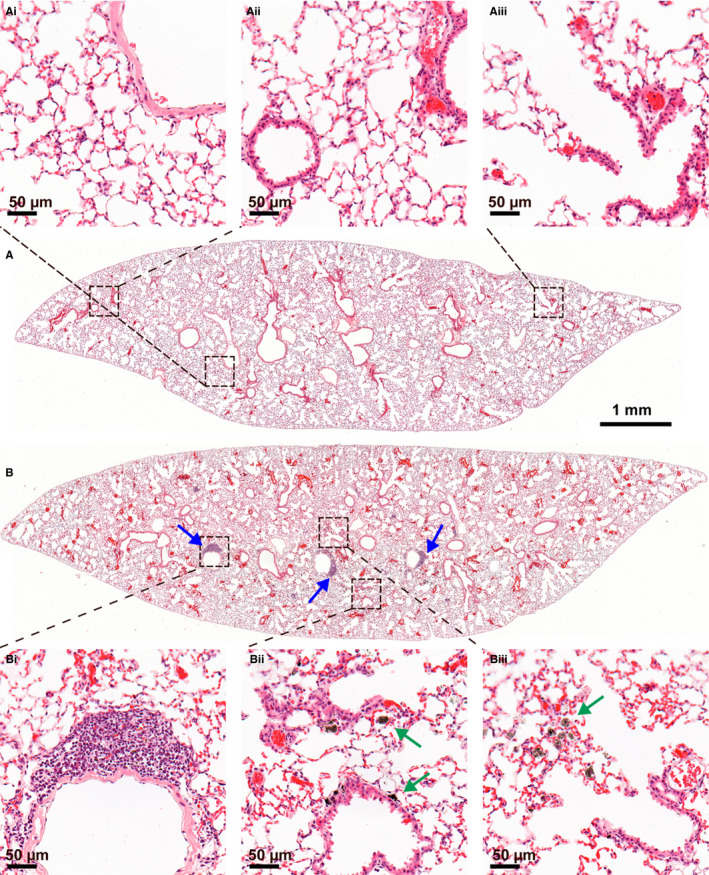
Multi-walled carbon nanotubes (MWCNT) are widely used nanomaterials that cause pulmonary toxicity upon inhalation. The physicochemical properties of MWCNT vary greatly, which makes general safety evaluation challenging to conduct. Identification of the toxicity-inducing physicochemical properties of MWCNT is therefore of great importance. We have evaluated histological changes in lung tissue 1 year after a single intratracheal instillation of 11 well-characterized MWCNT in female C57BL/6N BomTac mice. Genotoxicity in liver and spleen was evaluated by the comet assay. The dose of 54 μg MWCNT corresponds to three times the estimated dose accumulated during a work life at a NIOSH recommended exposure limit (0.001 mg/m3 ). Short and thin MWCNT were observed as agglomerates in lung tissue 1 year after exposure, whereas thicker and longer MWCNT were detected as single fibres, suggesting biopersistence of both types of MWCNT. The thin and entangled MWCNT induced varying degree of pulmonary inflammation, in terms of lymphocytic aggregates, granulomas and macrophage infiltration, whereas two thick and straight MWCNT did not. By multiple regression analysis, larger diameter and higher content of iron predicted less histopathological changes, whereas higher cobalt content significantly predicted more histopathological changes. No MWCNT-related fibrosis or tumours in the lungs or pleura was found. One thin and entangled MWCNT induced increased levels of DNA strand breaks in liver; however, no physicochemical properties could be related to genotoxicity. This study reveals physicochemical-dependent difference in MWCNT-induced long-term, pulmonary histopathological changes. Identification of diameter size and cobalt content as important for MWCNT toxicity provides clues for designing MWCNT, which cause reduced human health effects following pulmonary exposure.
Keywords: biodistribution; carbon nanotubes; granuloma; in vivo; lymphocytic aggregate; macrophage infiltration.
© 2018 The Authors. Basic & Clinical Pharmacology & Toxicology published by John Wiley & Sons Ltd on behalf of Nordic Association for the Publication of BCPT (former Nordic Pharmacological Society).
Conflict of interest statement
The authors report no conflict of interests.
Figures


Similar articles
-
Multi-walled carbon nanotube physicochemical properties predict pulmonary inflammation and genotoxicity.Nanotoxicology. 2016 Nov;10(9):1263-75. doi: 10.1080/17435390.2016.1202351. Epub 2016 Jul 7. Nanotoxicology. 2016. PMID: 27323647 Free PMC article.
-
Mouse pulmonary dose- and time course-responses induced by exposure to nitrogen-doped multi-walled carbon nanotubes.Inhal Toxicol. 2020 Jan;32(1):24-38. doi: 10.1080/08958378.2020.1723746. Epub 2020 Feb 7. Inhal Toxicol. 2020. PMID: 32028803 Free PMC article.
-
Mouse pulmonary dose- and time course-responses induced by exposure to multi-walled carbon nanotubes.Toxicology. 2010 Mar 10;269(2-3):136-47. doi: 10.1016/j.tox.2009.10.017. Epub 2009 Oct 24. Toxicology. 2010. PMID: 19857541
-
Inhalation toxicity assessment of carbon-based nanoparticles.Acc Chem Res. 2013 Mar 19;46(3):770-81. doi: 10.1021/ar200311b. Epub 2012 May 11. Acc Chem Res. 2013. PMID: 22574947 Review.
-
A review of toxicity studies of single-walled carbon nanotubes in laboratory animals.Regul Toxicol Pharmacol. 2016 Feb;74:42-63. doi: 10.1016/j.yrtph.2015.11.015. Epub 2015 Nov 24. Regul Toxicol Pharmacol. 2016. PMID: 26619783 Review.
Cited by
-
Effect of Surface Modification on the Pulmonary and Systemic Toxicity of Cellulose Nanofibrils.Biomacromolecules. 2022 Jul 11;23(7):2752-2766. doi: 10.1021/acs.biomac.2c00072. Epub 2022 Jun 9. Biomacromolecules. 2022. PMID: 35680128 Free PMC article.
-
Assessment of the Carcinogenicity of Carbon Nanotubes in the Respiratory System.Cancers (Basel). 2021 Mar 15;13(6):1318. doi: 10.3390/cancers13061318. Cancers (Basel). 2021. PMID: 33804168 Free PMC article. Review.
-
Continuous Long-Term Exposure to Low Concentrations of MWCNTs Induces an Epithelial-Mesenchymal Transition in BEAS-2B Cells.Nanomaterials (Basel). 2021 Jul 1;11(7):1742. doi: 10.3390/nano11071742. Nanomaterials (Basel). 2021. PMID: 34361127 Free PMC article.
-
Evaluation of total and inhalable samplers for the collection of carbon nanotube and carbon nanofiber aerosols.Aerosol Sci Technol. 2019;53(8):958-970. doi: 10.1080/02786826.2019.1618437. Epub 2019 May 30. Aerosol Sci Technol. 2019. PMID: 35392279 Free PMC article.
-
Pulmonary Toxicity of Long, Thick MWCNT and Very Long, Thin Carboxylated MWCNT Aerosols Following 28 Days Whole-Body Exposure.Toxics. 2025 May 16;13(5):401. doi: 10.3390/toxics13050401. Toxics. 2025. PMID: 40423481 Free PMC article.
References
-
- Jackson P, Kling K, Jensen KA, et al. Characterization of genotoxic response to 15 multiwalled carbon nanotubes with variable physicochemical properties including surface functionalizations in the FE1‐Muta(TM) mouse lung epithelial cell line. Environ Mol Mutagen. 2015;56:183‐203. - PubMed
-
- Beg S, Rizwan M, Sheikh AM, Hasnain MS, Anwer K, Kohli K. Advancement in carbon nanotubes: basics, biomedical applications and toxicity. J Pharm Pharmacol. 2011;63:141‐163. - PubMed
-
- Klumpp C, Kostarelos K, Prato M, Bianco A. Functionalized carbon nanotubes as emerging nanovectors for the delivery of therapeutics. Biochim Biophys Acta. 2006;1758:404‐412. - PubMed
MeSH terms
Substances
Grants and funding
LinkOut - more resources
Full Text Sources
Other Literature Sources
Medical
