

Peripheral Prisms Improve Obstacle Detection during Simulated Walking for Patients with Left Hemispatial Neglect and Hemianopia
- PMID: 30169355
- PMCID: PMC6260797
- DOI: 10.1097/OPX.0000000000001280
Peripheral Prisms Improve Obstacle Detection during Simulated Walking for Patients with Left Hemispatial Neglect and Hemianopia
Abstract
Significance: The first report on the use of peripheral prisms (p-prisms) for patients with left neglect and homonymous visual field defects (HVFDs).
Purpose: The purpose of this study was to investigate if patients with left hemispatial neglect and HVFDs benefit from p-prisms to expand the visual field and improve obstacle detection.
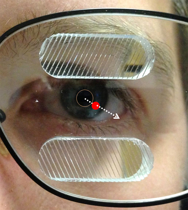
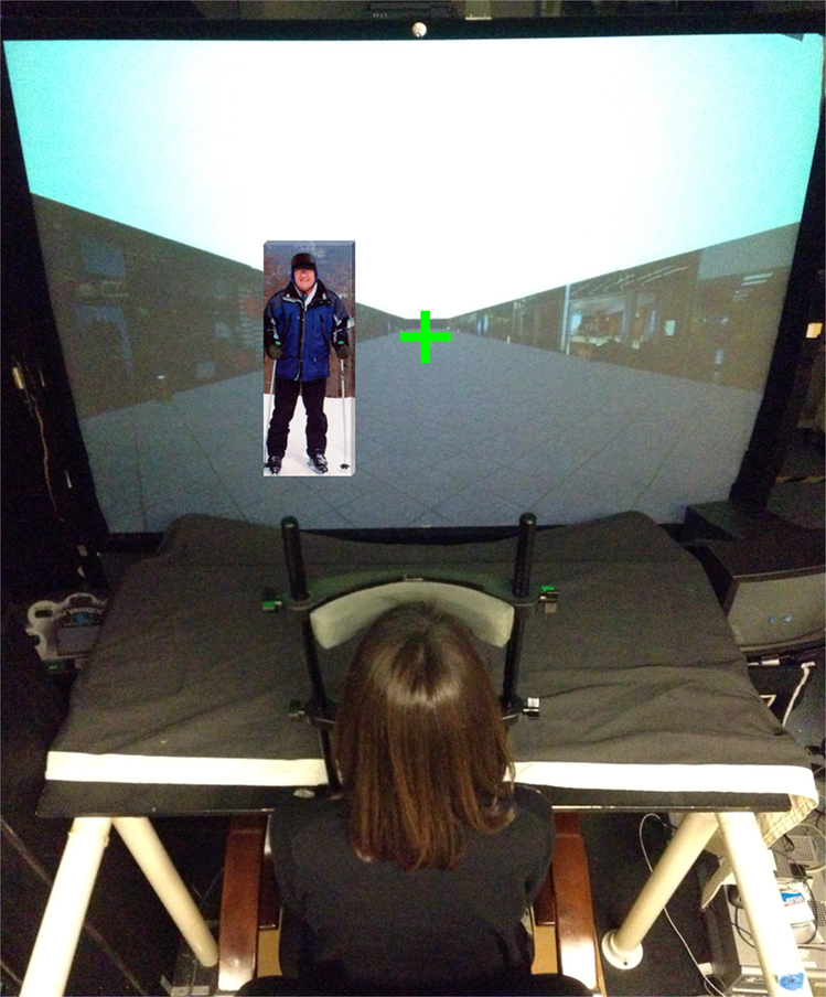
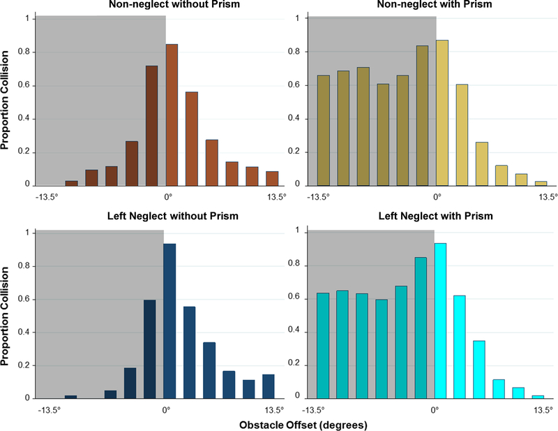
Methods: Patients (24 with HVFDs, 10 of whom had left neglect) viewed an animated, virtual, shopping mall corridor and reported if they would have collided with a human obstacle that appeared at various offsets up to 13.5° from their simulated walking path. There were 40 obstacle presentations on each side, with and without p-prisms. No training with p-prisms was provided, and gaze was fixed at the center of expansion.
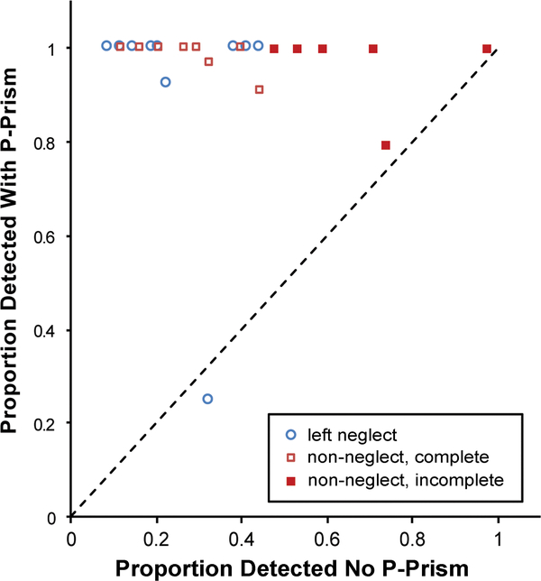
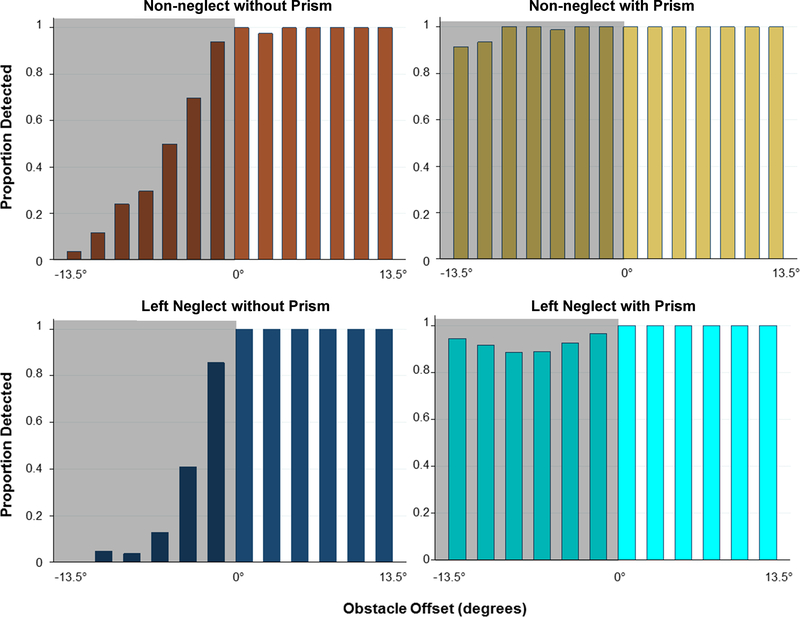
Results: Detection on the side of the HVFD improved significantly with p-prisms in both groups, from 26 to 92% in the left-neglect group and 43 to 98% in the non-neglect group (both P < .001). There was a tendency for greater improvement in the neglect patients with p-prisms. For collision judgments, both groups exhibited a large increase in perceived collisions on the side of the HVFD with the prisms (P < .001), with no difference between the groups (P = .93). Increased perceived collisions represent a wider perceived safety margin on the side of the HVFD.
Conclusions: Within the controlled conditions of this simulated, collision judgment task, patients with left neglect responded well to initial application of p-prisms exhibiting improved detection and wider safety margins on the side of the HVFD that did not differ from non-neglect patients. Further study of p-prisms for neglect patients in free-gaze conditions after extended wear and in real-world mobility tasks is clearly warranted.
Figures
Similar articles
-
Pilot study of a pedestrian collision detection test for a multisite trial of field expansion devices for hemianopia.Optom Vis Sci. 2024 Jun 1;101(6):408-416. doi: 10.1097/OPX.0000000000002152. Optom Vis Sci. 2024. PMID: 38990239 Free PMC article.
-
Effects of Perceptual-motor Training on Collision Judgments with Peripheral Prism Expanded Vision.Optom Vis Sci. 2022 Dec 1;99(12):875-884. doi: 10.1097/OPX.0000000000001957. Epub 2022 Nov 26. Optom Vis Sci. 2022. PMID: 36594755 Free PMC article.
-
Asymmetry in the Collision Judgments of People With Homonymous Field Defects and Left Hemispatial Neglect.Invest Ophthalmol Vis Sci. 2015 Jun;56(6):4135-42. doi: 10.1167/iovs.14-15492. Invest Ophthalmol Vis Sci. 2015. PMID: 26120818 Free PMC article.
-
Spontaneous recovery and treatment effects in patients with homonymous visual field defects: a meta-analysis of existing literature in terms of the ICF framework.Surv Ophthalmol. 2014 Jan-Feb;59(1):77-96. doi: 10.1016/j.survophthal.2013.02.006. Epub 2013 Oct 8. Surv Ophthalmol. 2014. PMID: 24112548 Review.
-
Object-centered visual neglect, or relative egocentric neglect?J Cogn Neurosci. 2000 May;12(3):542-5. doi: 10.1162/089892900562192. J Cogn Neurosci. 2000. PMID: 10931777 Review. No abstract available.
Cited by
-
Pilot study of a pedestrian collision detection test for a multisite trial of field expansion devices for hemianopia.Optom Vis Sci. 2024 Jun 1;101(6):408-416. doi: 10.1097/OPX.0000000000002152. Optom Vis Sci. 2024. PMID: 38990239 Free PMC article.
-
Toward Improving the Mobility of Patients with Peripheral Visual Field Defects with Novel Digital Spectacles.Am J Ophthalmol. 2020 Feb;210:136-145. doi: 10.1016/j.ajo.2019.10.005. Epub 2019 Oct 10. Am J Ophthalmol. 2020. PMID: 31606442 Free PMC article.
-
Gaze Scanning on Mid-Block Sidewalks by Pedestrians With Homonymous Hemianopia With or Without Spatial Neglect.Invest Ophthalmol Vis Sci. 2024 Jul 1;65(8):46. doi: 10.1167/iovs.65.8.46. Invest Ophthalmol Vis Sci. 2024. PMID: 39078731 Free PMC article.
-
Rehabilitation of visual perception in cortical blindness.Handb Clin Neurol. 2022;184:357-373. doi: 10.1016/B978-0-12-819410-2.00030-8. Handb Clin Neurol. 2022. PMID: 35034749 Free PMC article. Review.
-
Effects of Perceptual-motor Training on Collision Judgments with Peripheral Prism Expanded Vision.Optom Vis Sci. 2022 Dec 1;99(12):875-884. doi: 10.1097/OPX.0000000000001957. Epub 2022 Nov 26. Optom Vis Sci. 2022. PMID: 36594755 Free PMC article.
References
-
- Townend BS, Sturm JW, Petsoglou C, et al. Perimetric Homonymous Visual Field Loss Post-Stroke. J Clin Neurosci 2007;14:754–6. - PubMed
-
- Rowe F, Brand D, Jackson CA, et al. Visual Impairment Following Stroke: Do Stroke Patients Require Vision Assessment? Age Ageing 2009;38:188–93. - PubMed
-
- Zhang X, Kedar S, Lynn MJ, et al. Homonymous Hemianopias: Clinical-Anatomic Correlations in 904 Cases. Neurology 2006;66:906–10. - PubMed
-
- Schueller P, Micke O, Palkovic S, et al. 12 Years’ Experience with Intraoperative Radiotherapy (IORT) of Malignant Gliomas. Strahlenther Onkol 2005;181:500–6. - PubMed
-
- Kupersmith MJ, Vargas ME, Yashar A, et al. Occipital Arteriovenous Malformations: Visual Disturbances and Presentation. Neurology 1996;46:953–7. - PubMed
Publication types
MeSH terms
Grants and funding
LinkOut - more resources
Full Text Sources
Other Literature Sources
Medical
