Review
. 2018 Nov;34(6):413-419.
doi: 10.1097/MOG.0000000000000481.

On cell death in the intestinal epithelium and its impact on gut homeostasis

Affiliations
Review

On cell death in the intestinal epithelium and its impact on gut homeostasis

J Magarian Blander. Curr Opin Gastroenterol. 2018 Nov.

Abstract

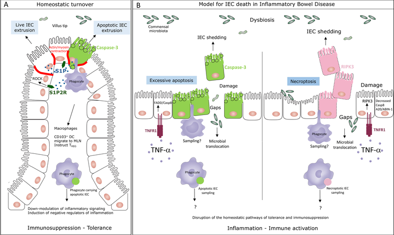
Purpose of review: Both apoptotic and nonapoptotic cell extrusion preserve the barrier functions of epithelia. Live cell extrusion is the paradigm for homeostatic renewal of intestinal epithelial cells (IEC). By extension, as extruded cells are not apoptotic, this form of cell shedding is thought to be largely ignored by lamina propria phagocytes and without immune consequence.

Recent findings: Visualization of apoptotic IEC inside distinct subsets of intestinal phagocytes during homeostasis has highlighted apoptosis as a normal component of the natural turnover of the intestinal epithelium. Analysis of phagocytes with or without apoptotic IEC corpses has shown how apoptotic IEC constrain inflammatory pathways within phagocytes and induce immunosuppressive regulatory CD4 T-cell differentiation. Many of the genes involved overlap with susceptibility genes for inflammatory bowel disease (IBD).

Summary: Excessive IEC death and loss-of-barrier function is characteristic of IBD. As regulatory and tolerogenic mechanisms are broken in IBD, a molecular understanding of the precise triggers and modes of IEC death as well as their consequences on intestinal inflammation is necessary. This characterization should guide new therapies that restore homeostatic apoptosis, along with its associated programs of immune tolerance and immunosuppression, to achieve mucosal healing and long-term remission.


Conflict of interest statement

Conflict of interest statement

None

Figures

Figure 1.
Figure 1.. The mode of cell death in the intestinal epithelium impacts intestinal homeostasis.
A. Under homeostatic conditions, the intestinal epithelium undergoes natural turnover by continuous replacement of intestinal epithelial cells (IEC). Experimental evidence supports both apoptotic and non-apoptotic IEC extrusion at locations of overcrowding at the villi tips of the small intestine or luminal face of the crypts in the colon. Both of these processes require sphingosine 1-phosphate (S1P) signaling through S1P2 receptor (S1P2R) and resultant Rho associated kinase (ROCK) dependent actin/myosin contraction. ROCK can be cleaved and activated by caspase-3 during apoptosis to phosphorylate myosin light chain and induce apoptotic membrane blebbing. MHC class II+ CD11c+ intestinal lamina propria phagocytes, either macrophages or CD103+CD11b dendritic cells (DC) sample the apoptotic IEC prior to their exclusion and respond by executing a broad program of immunosuppression. Apoptotic IEC carrying CD103+ DC also express negative regulators of inflammation such as the IBD susceptibility gene TNFAIP3 encoding A20. These cells express the lymph node homing receptor CCR7 and induce the emergence of regulatory CD4 T (TREG) cells. B. A proposed model for the types of IEC death that might occur during in inflammatory bowel disease. Increased levels of TNF-α in the intestinal lamina propria of patients with IBD induces IEC shedding that can be a result of excessive and unregulated apoptosis mediated by FADD and caspase-8 downstream of TNF receptor (TNFR1) engagement on IEC. Based on studies in mouse models, under conditions when caspase-8 or A20/ABIN-1 activity is impaired, TNF-α signaling through TNFR1 induces necroptosis mediated by the receptor interacting kinase 3 (RIPK3). Dying IEC are likely samples by intestinal phagocytes although formal proof of this process and its consequences is yet to come. In the absence of homeostatic apoptosis, it is expected that the immunosuppression and tolerance program in phagocytes, imparted by apoptotic IEC sampling, would be disrupted. Necroptosis is inflammatory in nature and would be expected to lead to the activation of intestinal phagocytes. Excessive apoptosis and necroptosis would compromise barrier integrity and lead to the translocation of the commensal microbiota into the sterile lamina propria leading to more inflammation.

References

    1. Ungaro R, Mehandru S, Allen PB, Peyrin-Biroulet L, Colombel JF. Ulcerative colitis. Lancet 2017;389(10080):1756–70. - PMC - PubMed
    1. Torres J, Mehandru S, Colombel JF, Peyrin-Biroulet L. Crohn’s disease. Lancet 2017;389(10080):1741–55. - PubMed
    1. Ramachandran A, Madesh M, Balasubramanian KA. Apoptosis in the intestinal epithelium: its relevance in normal and pathophysiological conditions. Journal of gastroenterology and hepatology 2000;15(2):109–20. - PubMed
    1. Nunes T, Bernardazzi C, de Souza HS. Cell death and inflammatory bowel diseases: apoptosis, necrosis, and autophagy in the intestinal epithelium. BioMed research international 2014;2014:218493. - PMC - PubMed
    1. Di Sabatino A, Ciccocioppo R, Luinetti O, Ricevuti L, Morera R, Cifone MG, et al. Increased enterocyte apoptosis in inflamed areas of Crohn’s disease. Diseases of the colon and rectum 2003;46(11):1498–507. - PubMed

Publication types

MeSH terms