. 2018 Aug 31;13(8):e0203291.
doi: 10.1371/journal.pone.0203291. eCollection 2018.

Sequential super-resolution imaging using DNA strand displacement

Affiliations

Sequential super-resolution imaging using DNA strand displacement

Sandeep Pallikkuth et al. PLoS One. .

Abstract

Sequential labeling and imaging in fluorescence microscopy allows the imaging of multiple structures in the same cell using a single fluorophore species. In super-resolution applications, the optimal dye suited to the method can be chosen, the optical setup can be simpler and there are no chromatic aberrations between images of different structures. We describe a method based on DNA strand displacement that can be used to quickly and easily perform the labeling and removal of the fluorophores during each sequence. Site-specific tags are conjugated with unique and orthogonal single stranded DNA. Labeling for a particular structure is achieved by hybridization of antibody-bound DNA with a complimentary dye-labeled strand. After imaging, the dye is removed using toehold-mediated strand displacement, in which an invader strand competes off the dye-labeled strand than can be subsequently washed away. Labeling and removal of each DNA-species requires only a few minutes. We demonstrate the concept using sequential dSTORM super-resolution for multiplex imaging of subcellular structures.


Conflict of interest statement

The authors have declared that no competing interests exist.

Figures
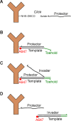
Fig 1
Fig 1. Concept of strand displacement.
(A) A protector strand is covalently conjugated to an antibody. (B) After labeling the cell with all antibodies of interest, a specific dye-labeled template strand is introduced to the buffer and binds to its matched protector strand. This sequence is long enough to prevent spontaneous dissociation. (C) After imaging, the template is removed by addition of an invader strand. The invader gains a toe-hold on the overhang section of the template and since individual base pairs can dissociate, the invader eventually out-competes the protector. (D) The dissociated double stranded invader/template is washed away.
Fig 2
Fig 2. Invader time course and residuals.
(Left) Set A. (Right) Set B. (A) Fluorescence intensity of a labeled cell sample as a function of time after invader addition. The cell is imaged for ~ 20 s before adding invader. (B) Super-resolution image before invader. (C) Super-resolution image after invader. The residual fluorescence for Set A and Set B are 2% and 5% respectively.
Fig 3
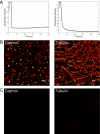
Fig 3. Sequential imaging results with clathrin and tubulin.
Clathrin and tubulin are labeled and imaged using Set A and Set B, respectively. (A) Super-resolution images acquired after 3 rounds of the label-image-remove process. (B) Overlay of all tubulin (cyan) and clathrin (yellow) imaging rounds. White box indicates regions shown in (A). (C) Comparison of the number of localization for each structure/set per imaging round.

References

    1. Dempsey G.T., Vaughan J.C., Chen K.H., Bates M., and Zhuang X.W., Evaluation of fluorophores for optimal performance in localization-based super-resolution imaging. Nature Methods, 2011. 8(12): p. 1027–+. 10.1038/nmeth.1768 - DOI - PMC - PubMed
    1. Tam J., Cordier G.A., Borbely J.S., Sandoval Álvarez Á., and Lakadamyali M., Cross-Talk-Free Multi-Color STORM Imaging Using a Single Fluorophore. PLoS ONE, 2014. 9(7): p. e101772 10.1371/journal.pone.0101772 - DOI - PMC - PubMed
    1. Rust M.J., Bates M., and Zhuang X.W., Sub-diffraction-limit imaging by stochastic optical reconstruction microscopy (STORM). Nature Methods, 2006. 3(10): p. 793–795. 10.1038/nmeth929 - DOI - PMC - PubMed
    1. Yi J., Manna A., Barr V.A., Hong J., Neuman K.C., and Samelson L.E., madSTORM: a superresolution technique for large-scale multiplexing at single-molecule accuracy. Molecular Biology of the Cell, 2016. 27(22): p. 3591–3600. 10.1091/mbc.E16-05-0330 - DOI - PMC - PubMed
    1. Heilemann M., van de Linde S., Schuttpelz M., Kasper R., Seefeldt B., Mukherjee A., Tinnefeld P., and Sauer M., Subdiffraction-resolution fluorescence imaging with conventional fluorescent probes. Angewandte Chemie-International Edition, 2008. 47(33): p. 6172–6176. 10.1002/anie.200802376 - DOI - PubMed

Publication types

LinkOut - more resources