. 2018 Dec 1;166(2):345-353.
doi: 10.1093/toxsci/kfy217.

Afghanistan Particulate Matter Enhances Pro-Inflammatory Responses in IL-13-Exposed Human Airway Epithelium via TLR2 Signaling

Affiliations

Afghanistan Particulate Matter Enhances Pro-Inflammatory Responses in IL-13-Exposed Human Airway Epithelium via TLR2 Signaling

Reena Berman et al. Toxicol Sci. .

Abstract

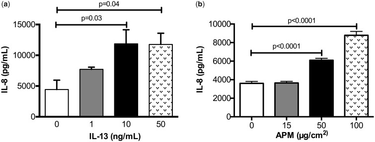
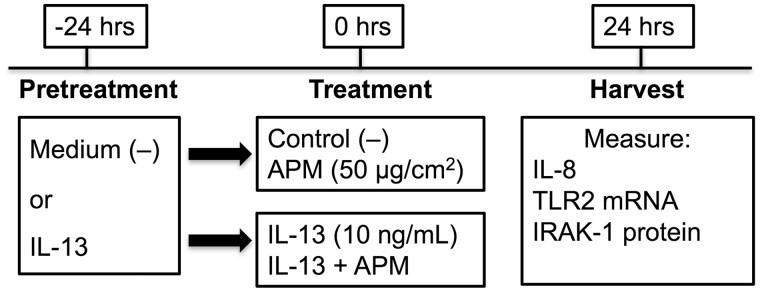
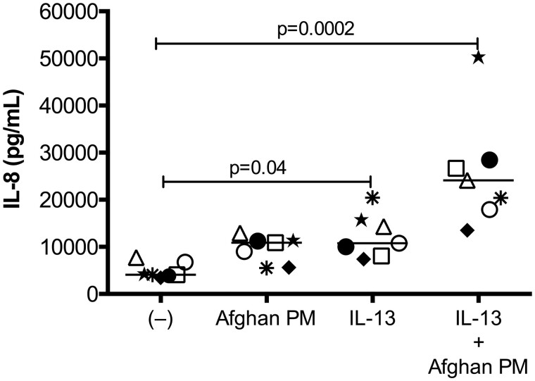
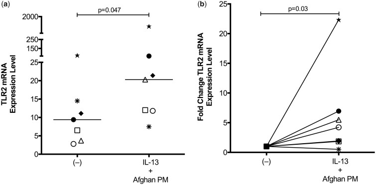
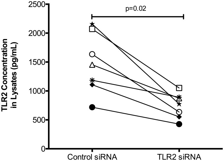
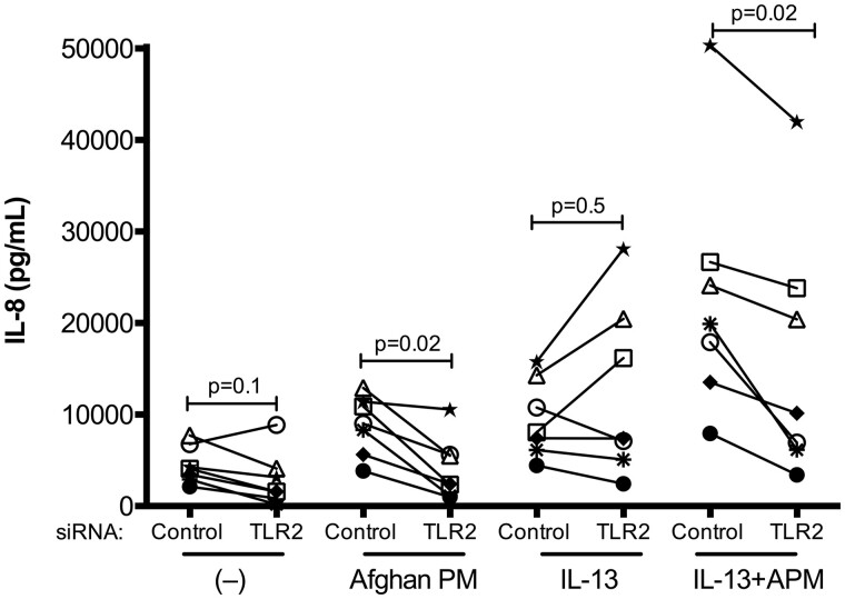
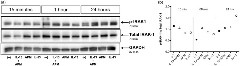
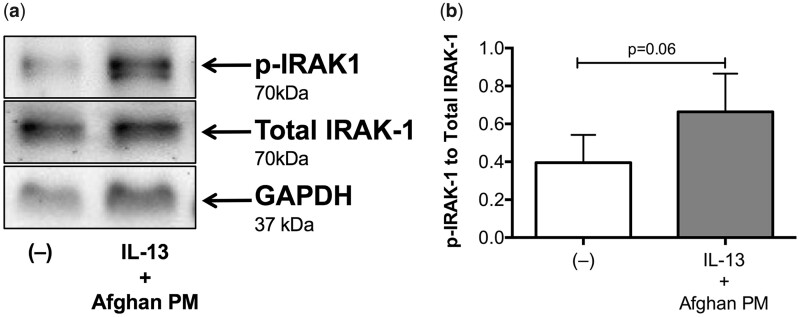
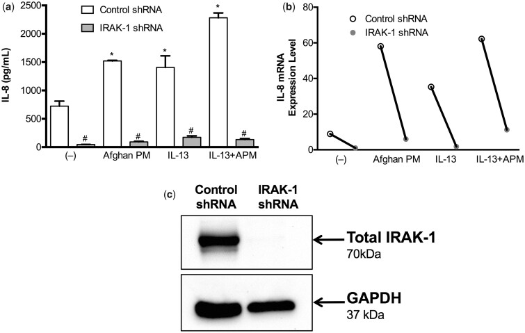
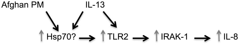
Since the start of Afghanistan combat operations in 2001, there has been an increase in complaints of respiratory illnesses in deployed soldiers with no previous history of lung disorders. It is postulated that deployment-related respiratory illnesses are the result of inhalation of desert particulate matter (PM) potentially acting in combination with exposure to other pro-inflammatory compounds. Why some, but not all, soldiers develop respiratory diseases remains unclear. Our goal was to investigate if human airway epithelial cells primed with IL-13, a type 2 inflammatory cytokine, demonstrate stronger pro-inflammatory responses to Afghanistan desert PM (APM). Primary human brushed bronchial epithelial cells from non-deployed, healthy subjects were exposed to APM, both with and without IL-13 pretreatment. APM exposure in conjunction with IL-13 resulted in significantly increased expression of IL-8, a pro-inflammatory cytokine involved in neutrophil recruitment and activation. Furthermore, expression of TLR2 mRNA was increased after combined IL-13 and APM exposure. siRNA-mediated TLR2 knockdown dampened IL-8 production after exposure to APM with IL-13. APM with IL-13 treatment increased IRAK-1 (a downstream signaling molecule of TLR2 signaling) activation, while IRAK-1 knockdown effectively eliminated the IL-8 response to APM and IL-13. Our data suggest that APM exposure may promote neutrophilic inflammation in airways with a type 2 cytokine milieu.


Figures

Figure 1.
Figure 1.
Size distribution and SEM image of Afghan particulate matter. Samples of Afghan PM from Bagram Air Force Base were analyzed for size distribution and particle shape via scanning electron microscope. Particles are classified as one of three sizes: Fine (<2.5 µm), Coarse (2.5–10 µm), and Large (>10 µm).
Figure 2.
Figure 2.
IL-13 and APM dose optimizations. Brushed bronchial cells from a normal subject (n = 1) were exposed to control (−) and IL−13 (1, 10, 50 ng/ml) (A) or control (−) and APM (15, 50, 100 ug/cm2) (B) and IL−8 was measured at each timepoint. A one-way ANOVA with Dunn’s multiple comparisons test was used to analyze differences between the treatment groups compared with the control (−) condition.
Figure 3.
Figure 3.
Overview of cell culture treatments. Schematic demonstrating cell culture conditions and treatments for in vitro APM exposures of bronchial epithelial cells.
Figure 4.
Figure 4.
Afghan PM induces IL-8 production in a type 2 inflammatory setting. Brushed bronchial cells from normal, non-deployed subjects (n = 7) were exposed to control (−), APM, IL-13, and the combination for 24 h. APM, IL-13, and the combination significantly increased IL-8 compared with medium control-treated cells. Each circle represents a unique patient and bars indicate the median. A paired Wilcoxon test was used to compare mean rank differences between two groups.
Figure 5.
Figure 5.
APM drives TLR2 mRNA expression in IL-13 exposed epithelial cells. Gene expression analysis of brushed bronchial cells from normal, non-deployed subjects (n = 7) exposed to control (−), APM, IL-13, and the combination for 24 h. Combination treatment of APM and IL-13 lead to significantly increased expression of TLR2 demonstrated via expression level (A) and fold change (B). Each circle represents a unique patient and bars indicate the median. A paired Wilcoxon test was used to compare mean rank differences between two groups.
Figure 6.
Figure 6.
Confirmation of TLR2 siRNA knockdown in cell lysates. Brushed bronchial cells from normal, non-deployed subjects (n = 7) were cultured under submerged conditions then transfected with either TLR2 siRNA or scrambled negative control siRNA. Cells were lysed and a TLR2 ELISA was used to confirm knockdown of the target gene. Each circle represents one unique subject. A paired Wilcoxon test was used to compare mean rank differences between groups.
Figure 7.
Figure 7.
Afghan PM-dependent IL-8 production is decreased after TLR2 knockdown. IL-8 ELISA of cells from non-deployed subjects (n = 7) transfected with negative control or TLR2 siRNA, then treated with control (−), APM, IL-13, and the combination. Each circle represents one unique subject. A paired Wilcoxon test was used to compare mean rank differences between control siRNA versus TLR2 siRNA within each treatment.
Figure 8.
Figure 8.
IRAK-1 activation occurs at the later phase after IL-13+APM treatment. Western blot analysis of bronchial epithelial cells from a non-deployed subject (Patient No. 2, n = 1) stimulated with control (−), APM, IL-13, and the combination for 15 min, 1 h, and 24 h (A). Duplicate wells were pooled at harvest in Western lysis buffer. Densitometry data with target proteins normalized to total protein (B).
Figure 9.
Figure 9.
Afghan PM and IL-13 signal through IRAK-1. Western blot analysis of bronchial epithelial cells from non-deployed subjects (n = 6) stimulated with control (−) or IL-13+APM. GAPDH was used as an internal loading control. Combination treatment of APM and IL-13 increases activation of p-IRAK1 (A). Densitometry data with target protein normalized to total protein levels (B).
Figure 10.
Figure 10.
IRAK-1 is essential for IL-8 production after APM and IL-13 treatment. Control and IRAK-1 shRNA was used to generate IRAK-1 knockdown tracheobronchial epithelial cells from one normal, deceased subject. After IRAK-1 shRNA, IL-8 production is significantly decreased across all conditions (A). IL-8 mRNA levels follow protein levels (B). Confirmation of IRAK-1 knockout via Western blot, with GAPDH as a loading control (C).
Figure 11.
Figure 11.
Overview of proposed APM and IL-13 signaling pathway.

References

    1. Army, U. S. Army Standards of Medical Fitness. Available at: https://www.calculator.net/pdf/r40_501.pdf. Accessed June 14 2017.
    1. Asea A., Rehli M., Kabingu E., Boch J. A., Bare O., Auron P. E., Stevenson M. A., Calderwood S. K. (2002). Novel signal transduction pathway utilized by extracellular HSP70: Role of toll-like receptor (TLR) 2 and TLR4. J. Biol. Chem. 277, 15028–15034. - PubMed
    1. Becker S., Dailey L., Soukup J. M., Silbajoris R., Devlin R. B. (2005). TLR-2 is involved in airway epithelial cell response to air pollution particles. Toxicol. Appl. Pharmacol. 203, 45–52. - PubMed
    1. Becker S., Fenton M. J., Soukup J. M. (2002). Involvement of microbial components and toll-like receptors 2 and 4 in cytokine responses to air pollution particles. Am. J. Respir. Cell Mol. Biol. 27, 611–618. - PubMed
    1. Breznan D., Karthikeyan S., Phaneuf M., Kumarathasan P., Cakmak S., Denison M. S., Brook J. R., Vincent R. (2016). Development of an integrated approach for comparison of in vitro and in vivo responses to particulate matter. Part. Fibre Toxicol. 13, 41. - PMC - PubMed

MeSH terms

LinkOut - more resources