. 2019 Jul 22;29(8):3266-3281.
doi: 10.1093/cercor/bhy194.

Adenosine Receptor-Mediated Developmental Loss of Spike Timing-Dependent Depression in the Hippocampus

Affiliations

Adenosine Receptor-Mediated Developmental Loss of Spike Timing-Dependent Depression in the Hippocampus

Mikel Pérez-Rodríguez et al. Cereb Cortex. .

Abstract

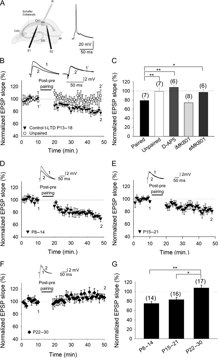
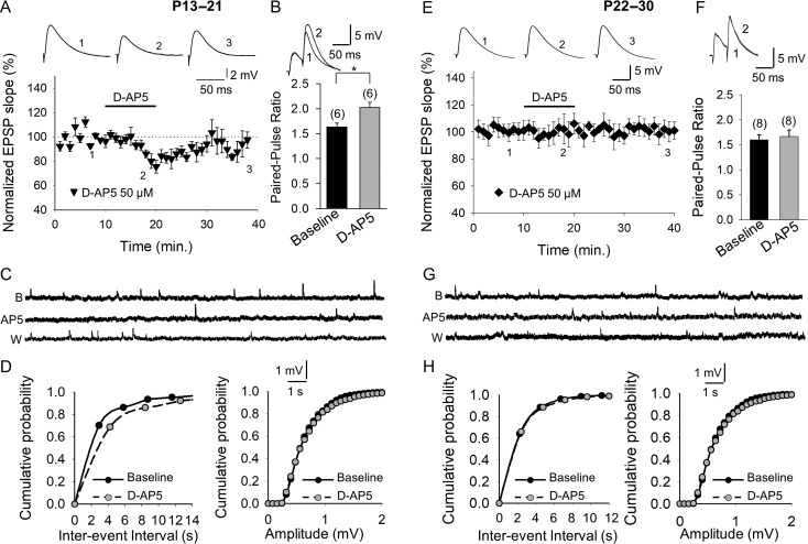
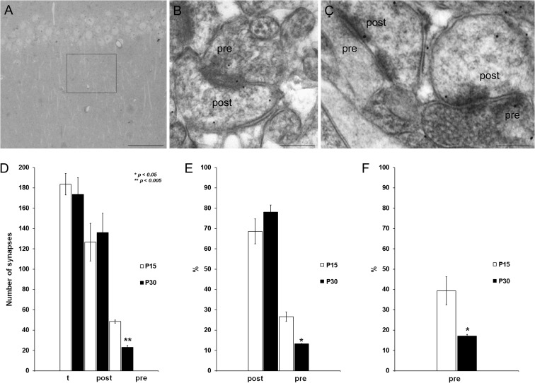
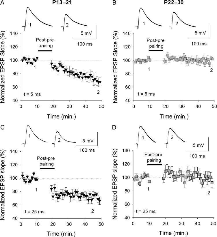
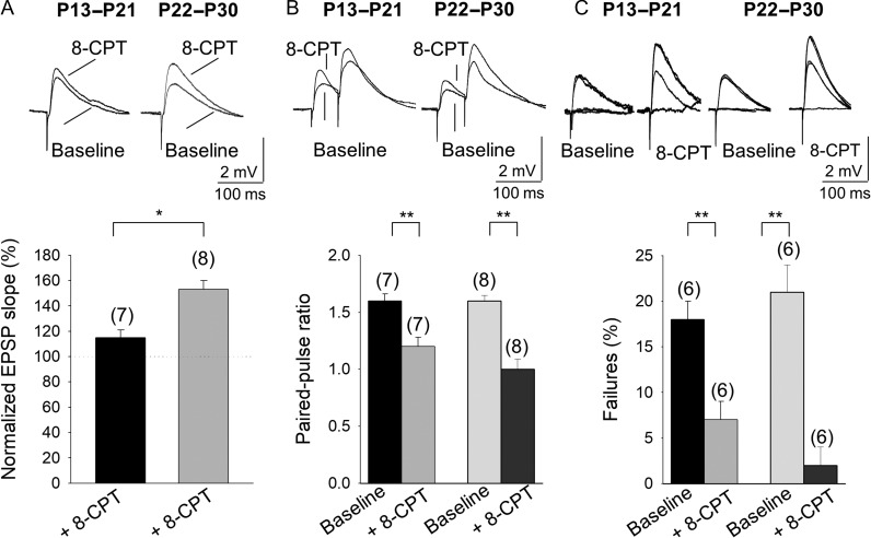
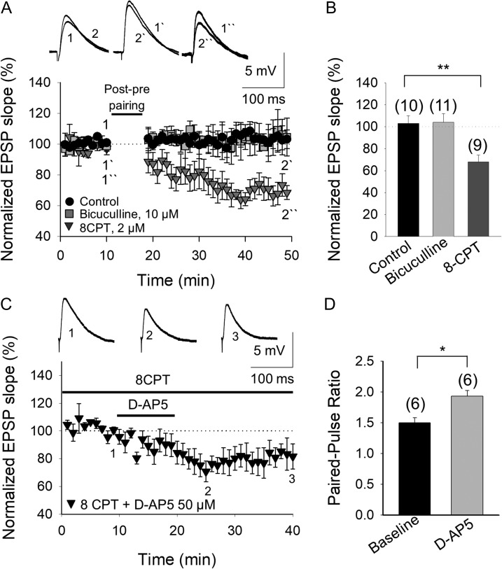
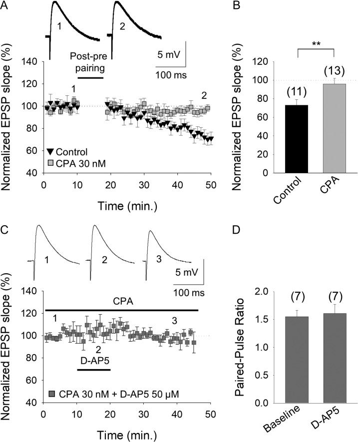
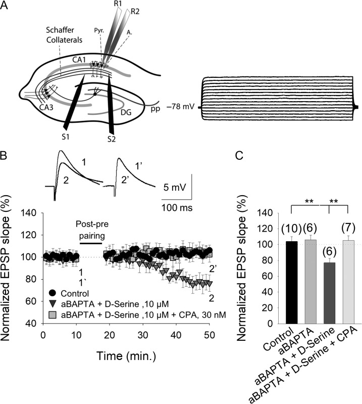
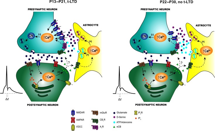
Critical periods of synaptic plasticity facilitate the reordering and refining of neural connections during development, allowing the definitive synaptic circuits responsible for correct adult physiology to be established. Presynaptic spike timing-dependent long-term depression (t-LTD) exists in the hippocampus, which depends on the activation of NMDARs and that probably fulfills a role in synaptic refinement. This t-LTD is present until the third postnatal week in mice, disappearing in the fourth week of postnatal development. We were interested in the mechanisms underlying this maturation related loss of t-LTD and we found that at CA3-CA1 synapses, presynaptic NMDA receptors (pre-NMDARs) are tonically active between P13 and P21, mediating an increase in glutamate release during this critical period of plasticity. Conversely, at the end of this critical period (P22-P30) and coinciding with the loss of t-LTD, these pre-NMDARs are no longer tonically active. Using immunogold electron microscopy, we demonstrated the existence of pre-NMDARs at Schaffer collateral synaptic boutons, where a decrease in the number of pre-NMDARs during development coincides with the loss of both tonic pre-NMDAR activation and t-LTD. Interestingly, this t-LTD can be completely recovered by antagonizing adenosine type 1 receptors (A1R), which also recovers the tonic activation of pre-NMDARs at P22-P30. By contrast, the induction of t-LTD was prevented at P13-P21 by an agonist of A1R, as was tonic pre-NMDAR activation. Furthermore, we found that the adenosine that mediated the loss of t-LTD during the fourth week of development is supplied by astrocytes. These results provide direct evidence for the mechanism that closes the window of plasticity associated with t-LTD, revealing novel events probably involved in synaptic remodeling during development.

Keywords: adenosine receptors; astrocytes; hippocampus; plasticity windows; spike timing-dependent plasticity.


Figures

Figure 1.
Figure 1.
Input-specific spike timing-dependent plasticity in the CA1 region of the hippocampus is present at P8–P14 and P15–P21 but not at P22–P30. (A) Left, scheme showing the general experimental set-up: R, recording electrode; S1 and S2, stimulating electrodes; right, pairing protocol utilized (Δt, time between EPSP onset and peak of spike). (B) Post–pre single-spike pairing protocol induced t-LTD. The EPSP slopes monitored in the paired (black circles) and unpaired pathway (open circles) are shown. Traces show the EPSP before (1) and 30 min after (2) pairing. Depression was only observed in the paired pathway. (C) Summary of the results. Note that t-LTD does not require postsynaptic NMDARs. In the presence of d-AP5 (50 μM), t-LTD was completely blocked with MK-801 added to the bath (eMK-801), yet t-LTD was not affected when MK-801 was loaded into the postsynaptic cell (iMK-801). The t-LTD is evident during the second (D) and third (E) week of development but it disappears during the fourth week (F). (G) Summary of the results, where the error bars represent the S.E.M. and the number of slices is shown in parentheses: *P < 0.05; **P < 0.01, unpaired Student’s t-test.
Figure 2.
Figure 2.
Presynaptic NMDARs are tonically active at CA3–CA1 synapses at P13–P21 but not at P22–P30. (A) With the postsynaptic neuron loaded with MK-801, the addition of D-AP5 decreases the slope of evoked EPSPs, an effect that was reversed after D-AP5 washout. The inset shows the EPSP traces at baseline (1), in the presence of D-AP5 (2) and after D-AP5 wash out (3). (B) The paired-pulse ratio increased in the presence of D-AP5. (C) Miniature EPSPs monitored during the baseline and after exposing neurons from slices of P13–P21 animals to D-AP5 in the presence of TTX (500 nM), and with the postsynaptic neuron loaded with MK-801 (1 mM). (D) Cumulative probability histograms showing that at P13–P21, D-AP5 reversibly decreases the mEPSP frequency but it does not affect the mEPSP amplitude. (E) D-AP5 does not affect the evoked EPSP slope at P22–P30. The inset shows the traces at baseline (1), in the presence of D-AP5 (2) and after D-AP5 wash out (3). (F) At P22–P30, the paired-pulse ratio is not affected by D-AP5. (G) Miniature EPSPs monitored at baseline, and during and after D-AP5 treatment of neurons from P22 to P30 mice in the presence of TTX, and with the postsynaptic neuron loaded with MK-801. (H) Cumulative probability histograms showing that D-AP5 does not affect mEPSP frequency or amplitude at P22–P30. The error bars indicate the S.E.M. and the number of slices is shown in parentheses: *P < 0.05, unpaired Student’s t-test.
Figure 3.
Figure 3.
Presynaptic NR1 is downregulated during development. (A) Transmitted light photomicrograph illustrating the CA1 area of the mouse hippocampus analyzed (square). (B, C) Electron photomicrographs of the CA1 demonstrating the presence of NR1 immunolabeling (arrows) at both presynaptic (pre) and postsynaptic (post) sites at P15 (B) and P30 (C). Scale bars: 200 μm (A) and 200 nm (B, C). (D) The number of NR1 immunolabeled postsynaptic sites did not change from P15 to P30, whereas the number of immunolabeled presynaptic terminals decreased significantly at P30 compared with P15. (E) The proportion of presynaptic labeled terminals relative to the total number of synapses decreases with age, while the percentage of labeled postsynaptic terminals does not change significantly. (F) A statistically significant decrease was also observed when the proportion of presynaptic labeled terminals was compared with the number of NR1 labeled postsynaptic terminals: t, total number of synapses counted, n = 4 mice (2 for each age analyzed). The error bars represent the S.E.M., and >150 synapses per mouse and region were analyzed: *P < 0.05; **P < 0.01, unpaired Student’s t-test.
Figure 4.
Figure 4.
The loss of t-LTD is not due to a shift in the coincidence time window to induce t-LTD. Using timings between presynaptic and postsynaptic activity to induce t-LTD that were shorter and longer than 18 ms (5 and 25 ms), t-LTD is evident at P13–P21 and lost at P22–P30, both at the 5 and 25 ms intervals, as occurred with an interval of 18 ms: (A) P13–P21 t = 5 ms; (B) P22–P30, t = 5 ms; (C) P13–P21, t = 25 ms; (D) P22–P30, t = 25 ms.
Figure 5.
Figure 5.
Presynaptic A1R-mediated inhibition increases with maturation. (A) 8-CPT affects the evoked EPSP slope at P13–P21 and P22–P30. (B) Effect of 8-CPT on the paired-pulse ratio (PPR). Note that 8-CPT produces a decrease in the PPR at P13–P21 and P22–P30. (C) The effect of 8-CPT on the number of failures of synaptic transmission. Note that 8-CPT produces a decrease in the number of failures. Error bars reflect the S.E.M. and the number of slices is shown in parentheses: *P < 0.05; **P < 0.01, unpaired Student’s t-test.
Figure 6.
Figure 6.
The developmental loss of t-LTD involves an increase in the inhibition mediated by adenosine A1 type receptor activation. (A) The loss of t-LTD is due to the activation of A1Rs and not to the activation of GABAA receptors. The t-LTD lost at P22–P30 was not recovered in the presence of bicuculline (grey squares), whereas the lost t-LTD is completely recovered in the presence of the A1R antagonist 8-CPT (2 μM, grey triangles). The insets show the EPSP before (1, 1´, 1´´) and after (2, 2´, 2´´) post–pre pairing in control conditions (black circles), in the presence of bicuculline or 8-CPT. (B) Summary of the results. (C) The tonic activation of pre-NMDARs that is lost at P22–P30 is completely recovered when A1Rs are antagonized. In the presence of 8-CPT and with the postsynaptic neuron loaded with MK-801, D-AP5 induces a reversible decrease in the EPSP slope. Traces show the EPSP before (1), during (2), and after (3) exposure to D-AP5. (D) The paired-pulse ratio increases in the presence of D-AP5. The error bars indicate the S.E.M. and the number of slices is shown in parentheses: *P < 0.05; **P < 0.01, unpaired Student’s t-test.
Figure 7.
Figure 7.
An increase in A1R mediated-inhibition closes the window of plasticity for t-LTD at P13–P21. (A) Activation of A1Rs at P13–P21 by the agonist CPA prevented the induction of t-LTD. The evoked EPSP slopes monitored in control slices (black triangles) and in slices treated with the A1R agonist CPA (grey squares) following a post–pre pairing are shown. The traces show the EPSP before (1, 1´) and 30 min after (2, 2´) pairing in control slices, and in slices treated with CPA. (B) Summary of the results. (C) Tonic pre-NMDAR activation at P13–P21 is lost in the presence of the A1R agonist CPA. D-AP5 did not affect the evoked EPSP slope in the presence of CPA. The traces show the EPSP before 1), during 2), and after 3) exposure to D-AP5. (D) The paired-pulse ratio was not affected by D-AP5 in the presence of CPA (with the postsynaptic cell loaded with MK-801). The error bars represent the S.E.M. and the number of slices is shown in parentheses: **P < 0.01, unpaired Student’s t-test.
Figure 8.
Figure 8.
The adenosine involved in preventing t-LTD at P22–P30 is from astrocytes. (A) Left, scheme showing the general experimental set-up: R1 and R2, recording electrodes; S1 and S2, stimulating electrodes; Pyr, pyramidal neuron; A, astrocyte; right, voltage responses of an astrocyte shown in current-clamp. (B) In astrocyte-neuron dual recordings, with the calcium chelator BAPTA injected into the astrocyte via the recording pipette (aBAPTA), and with D-serine (100 μM) added to the bath, a post–pre pairing protocol induced t-LTD (grey triangles) but not in control conditions (no BAPTA and no d-serine, black circles). The presence of CPA impaired the t-LTD observed with aBAPTA and d-serine (dark grey squares). Inset: representative traces at the baseline (1 and 1´) and 30 min after the pairing protocol (2 and 2´) in the presence of aBAPTA and d-serine alone, with or without CPA. (C) Summary of the results, with the error bars reflecting the S.E.M. and the number of slices shown in parentheses: **P < 0.01, unpaired Student’s t-test.
Figure 9.
Figure 9.
Scheme of the differences in signaling between early (P13–P21) and late (P22–P30) stages of development. (A) At P13–P21, t-LTD is induced by a post–pre single-spike pairing protocol. Postsynaptic action potentials activate voltage-dependent Ca2+ channels (VDCCs) and the presynaptically released glutamate activates postsynaptic mGlu5 receptors. These receptors synergistically activate PLC and produce IP3, provoking Ca2+ release from internal stores and DAG production, which serves as a precursor for endocannabinoid (eCB) synthesis. The eCB signal activates CB1 receptors, facilitating d-serine release from astrocytes. Together with the glutamate released from presynaptic neurons, this d-serine activates presynaptic NMDARs on Schaffer Collateral boutons. This leads to an increase in presynaptic Ca2+, calcineurin activation and synaptic depression. (B) At P22–P30, t-LTD does not develop and the main differences at these synapses at the 2 stages of development are: a change in the probability of glutamate release (higher at P13–P21 than at P22–P30), the tonic activation of pre-NMDARs at P13–P21 but not at P22–P30, a decrease in the number of pre-NMDARs at P22–P30, and an increase in adenosine release from astrocytes at P22–P30 compared with P13–21.

Similar articles

Cited by

References

    1. Andrade-Talavera Y, Duque-Feria P, Negrete-Díaz JV, Sihra TS, Flores G, Rodríguez-Moreno A. 2012. Presynaptic kainate receptor-mediated facilitation of glutamate release involves Ca2+-calmodulin at mossy fiber-CA3 synapses. J Neurochem. 122:891–899. - PubMed
    1. Andrade-Talavera Y, Duque-Feria P, Paulsen O, Rodríguez-Moreno A. 2016. Presynaptic spike timing-dependent long-term depression in the mouse hippocampus. Cereb Cortex. 26:3637–3654. - PMC - PubMed
    1. Arai A, Kessler M, Lynch G. 1990. The effects of adenosine on the development of long-term potentiation. Neurosci Lett. 119:41–44. - PubMed
    1. Araque A, Carmignoto G, Haydon PG, Oliet SH, Robitaille R, Volterra A. 2014. Gliotransmitters travel in time and space. Neuron. 81:728–739. - PMC - PubMed
    1. Banerjee A, Meredith RM, Rodríguez-Moreno A, Mierau SB, Auberson YP, Paulsen O. 2009. Double dissociation of spike timing-dependent potentiation and depression by subunit-preferring NMDA receptors antagonists in mouse barrel cortex. Cereb Cortex. 19:2959–2969. - PMC - PubMed

MeSH terms