

Granular Cell Tumor of the Bladder: A Report of Six Cases
- PMID: 30170086
- PMCID: PMC8043247
- DOI: 10.1016/j.urology.2018.08.018
Granular Cell Tumor of the Bladder: A Report of Six Cases
Abstract
Objective: To better characterize granular cell tumor of the bladder, with only 20 cases reported to date and unclear management guidelines.
Methods: We report five benign and one malignant granular cell tumor of the bladder.
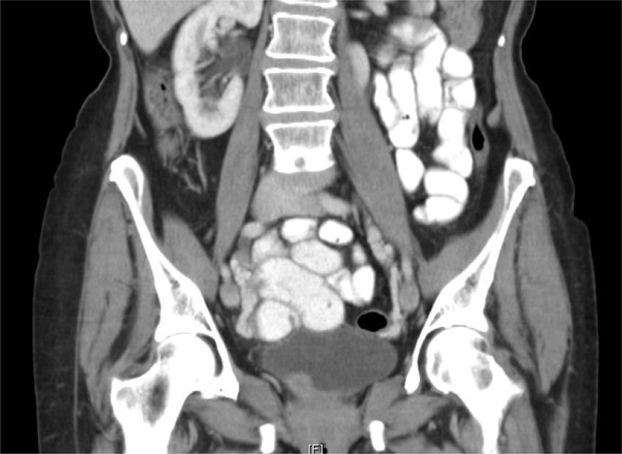
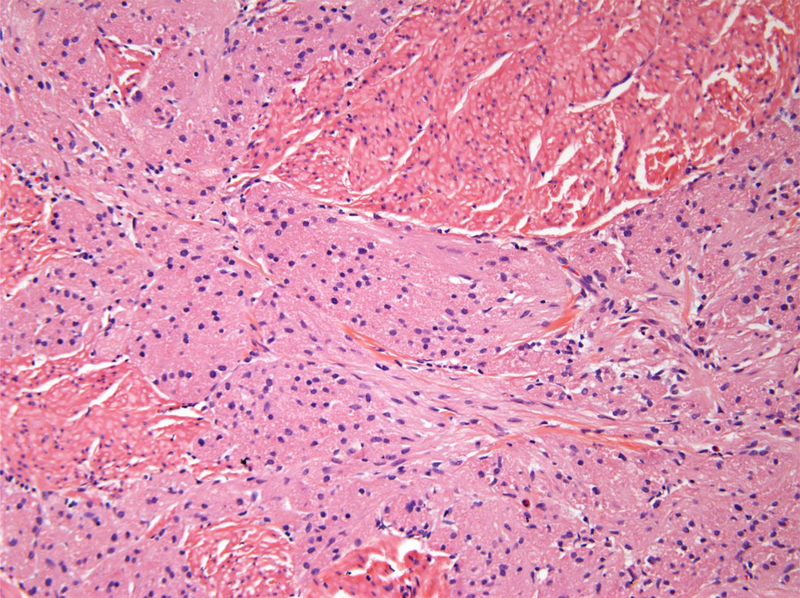
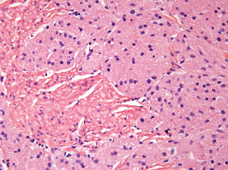
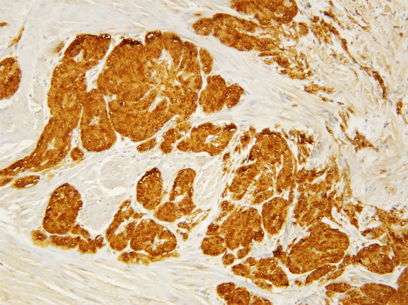
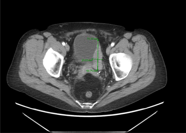
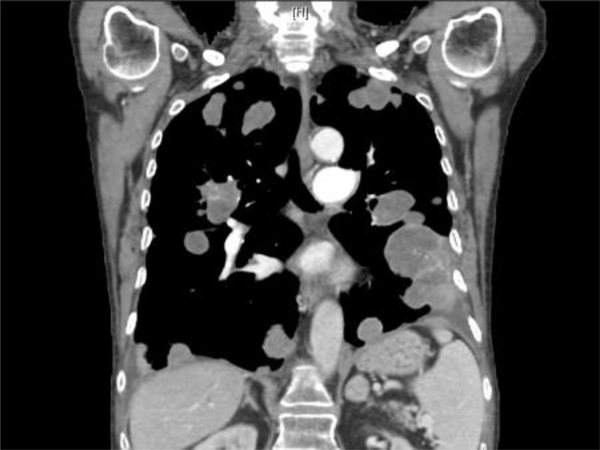
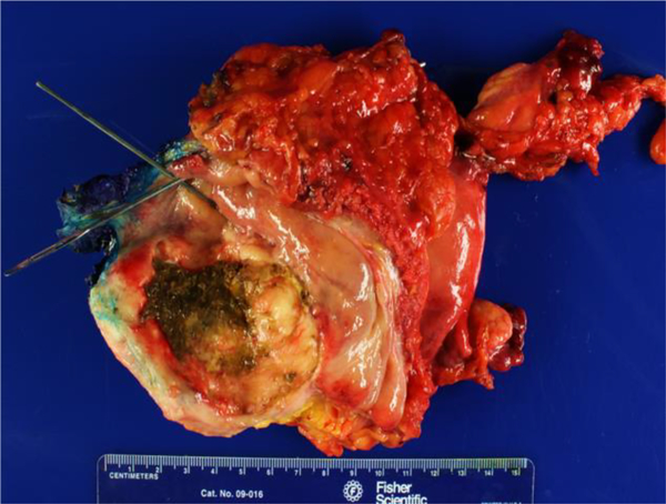
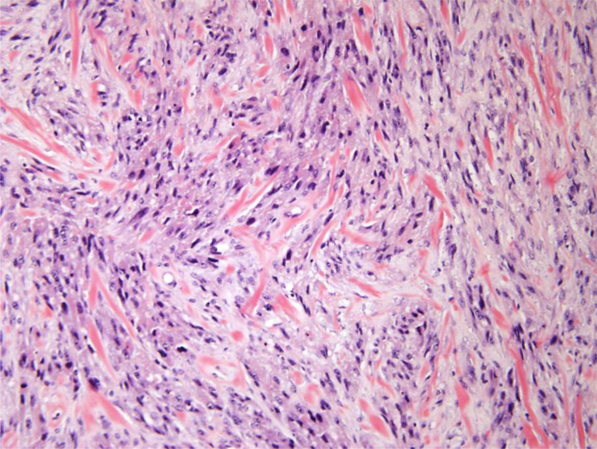
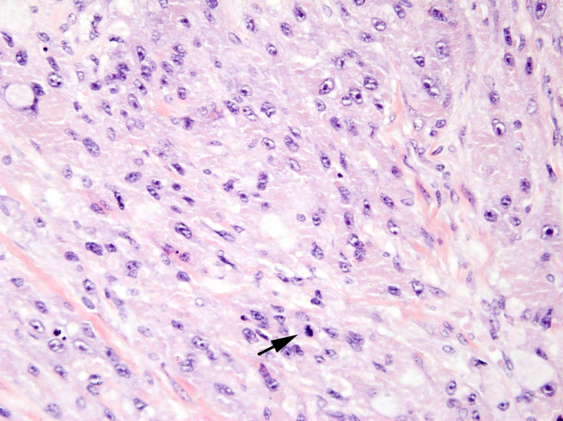
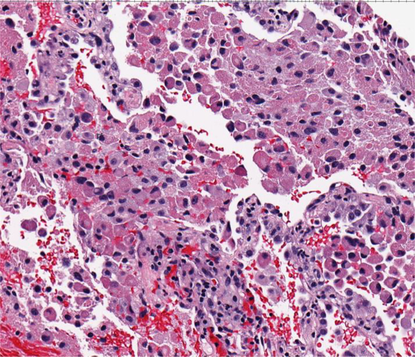
Results: Patients were in the age range of 33 to 73 years. The size of tumor ranged from 0.2 to 6.3 cm. Two benign granular cell tumors were incidental findings with others presenting with painless hematuria. Benign granular cell tumors infiltrated the muscularis propria and were confirmed by immunohistochemistry for S100 protein with negative stains for keratins. The malignant granular cell tumor involved the entire bladder wall with extension into perivesical tissue. Benign granular cell tumors were treated by transurethral resection (TUR) or partial cystectomy; all patients were disease free at last follow-up. The malignant granular cell tumor was treated by anterior exenteration and bilateral pelvic lymphadenectomy. This patient developed pulmonary and pleural metastases 2 years after surgery.
Conclusion: Given the locally infiltrative nature of granular cell tumors and that 50% of reported benign granular cell tumors with sufficient follow-up recurred following initial TUR, it is prudent to recommend partial cystectomy if technically feasible. A later TUR at a time of tumor regrowth could result in obstruction of ureters depending on their location and with greater infiltrative growth, with larger subsequent resections be needed for complete removal. In other cases, immediate repeat TUR after a diagnosis of granular cell tumor would lessen the likelihood of local recurrence. Either partial or radical cystectomy is needed for the rare malignant granular cell tumor.
Copyright © 2018 Elsevier Inc. All rights reserved.
Figures
Similar articles
-
Granular cell tumors of the urinary bladder.World J Surg Oncol. 2007 Mar 13;5:33. doi: 10.1186/1477-7819-5-33. World J Surg Oncol. 2007. PMID: 17355632 Free PMC article.
-
[Non-epithelial tissue tumors of the urinary bladder].Zhonghua Wai Ke Za Zhi. 2003 Jul;41(7):530-3. Zhonghua Wai Ke Za Zhi. 2003. PMID: 12921662 Chinese.
-
Inflammatory pseudotumor and sarcoma of urinary bladder: differential diagnosis and outcome in thirty-eight spindle cell neoplasms.Mod Pathol. 2001 Oct;14(10):1043-51. doi: 10.1038/modpathol.3880434. Mod Pathol. 2001. PMID: 11598176
-
Challenges in Pathologic Staging of Bladder Cancer: Proposals for Fresh Approaches of Assessing Pathologic Stage in Light of Recent Studies and Observations Pertaining to Bladder Histoanatomic Variances.Adv Anat Pathol. 2017 May;24(3):113-127. doi: 10.1097/PAP.0000000000000152. Adv Anat Pathol. 2017. PMID: 28398951 Review.
-
[Analysis of 34 cases of infiltrating carcinoma of the bladder treated exclusively with partial cystectomy (part 1)].Arch Esp Urol. 1996 May;49(4):349-64. Arch Esp Urol. 1996. PMID: 8754191 Review. Spanish.
Cited by
-
A case report of rare granular cell tumor of the urinary bladder.Urol Case Rep. 2022 Feb 16;42:102034. doi: 10.1016/j.eucr.2022.102034. eCollection 2022 May. Urol Case Rep. 2022. PMID: 35530539 Free PMC article.
-
Atypical granular cell tumour of urinary bladder - case report of an extremely rare neoplasm.Cent European J Urol. 2024;77(4):637-640. doi: 10.5173/ceju.2024.153. Epub 2024 Nov 10. Cent European J Urol. 2024. PMID: 40313708 Free PMC article.
-
Atypical granular cell tumor of the urinary bladder: A case report.World J Clin Cases. 2021 Oct 6;9(28):8453-8460. doi: 10.12998/wjcc.v9.i28.8453. World J Clin Cases. 2021. PMID: 34754853 Free PMC article.
References
-
- Abrikossoff A. Uber Myome ausgehend von der quergestreiften Willkurlichen muskulatur. Virchows Arch Pathol Anal 1926; 260: 215–33.
-
- Fanburg-Smith JC, Meis-Kindblom JM, Fante R, et al. Malignant granular cell tumor of soft tissue: diagnostic criteria and clinicopathologic correlation. Am J Surg Pathol 1998; 22: 779–94. - PubMed
-
- Andersen R, Hoeg K. Myoblastoma of the bladder neck: report of a case. Br J Urol 1961; 33: 76–9. - PubMed
Publication types
MeSH terms
Grants and funding
LinkOut - more resources
Full Text Sources
Other Literature Sources
Medical
