Review
. 2019 Apr;143(4):1271-1286.
doi: 10.1016/j.jaci.2018.07.037. Epub 2018 Aug 29.

Airways exudation of plasma macromolecules: Innate defense, epithelial regeneration, and asthma

Affiliations
Review

Airways exudation of plasma macromolecules: Innate defense, epithelial regeneration, and asthma

Carl Persson. J Allergy Clin Immunol. 2019 Apr.

Abstract

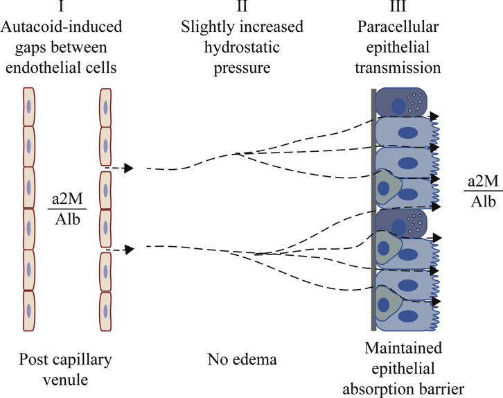
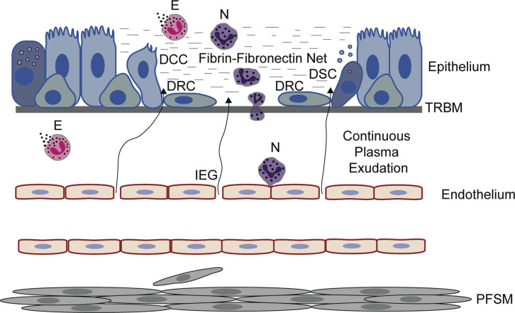
This review discusses in vivo airway aspects of plasma exudation in relation to current views on epithelial permeability and epithelial regeneration in health and disease. Microvascular-epithelial exudation of bulk plasma proteins characteristically occurs in asthmatic patients, being especially pronounced in those with severe and exacerbating asthma. Healthy human and guinea pig airways challenged by noninjurious histamine-leukotriene-type autacoids also respond through prompt mucosal exudation of nonsieved plasma macromolecules. Contrary to current beliefs, epithelial permeability in the opposite direction (ie, absorption of inhaled molecules) has not been increased in patients with asthma and allergic rhinitis or in acutely exuding healthy airways. A slightly increased subepithelial hydrostatic pressure produces such unidirectional outward perviousness to macromolecules. Lack of increased absorption permeability in asthmatic patients can further be reconciled with occurrence of epithelial shedding, leaving small patches of denuded basement membrane. Counteracting escalating barrier breaks, plasma exudation promptly covers the denuded patches. Here it creates and sustains a biologically active barrier involving a neutrophil-rich, fibrin-fibronectin net. Furthermore, in the plasma-derived milieu, all epithelial cell types bordering the denuded patch dedifferentiate and migrate from all sides to cover the denuded basement membrane. However, this speedy epithelial regeneration can come at a cost. Guinea pig in vivo studies demonstrate that patches of epithelial denudation regeneration are exudation hot spots evoking asthma-like features, including recruitment/activation of granulocytes, proliferation of fibrocytes/smooth muscle cells, and basement membrane thickening. In conclusion, nonsieved plasma macromolecules can operate on the intact airway mucosa as potent components of first-line innate immunity responses. Exuded plasma also takes center stage in epithelial regeneration. When exaggerated, epithelial regeneration can contribute to the inception and development of asthma.

Keywords: Plasma proteins; airway epithelium; airway microcirculation; asthma pathogenesis; epithelial permeability; epithelial regeneration; innate immunity.


Figures

Fig 1
Fig 1
The 3 major steps involved in airway microvascular-epithelial exudation of nonsieved plasma proteins. After local mucosal challenge, autacoids induce high-permeability gaps between endothelial cells in postcapillary venules (I). Venules belong to a profuse subepithelial microcirculation distinct from the pulmonary circulation. Through these gaps, plasma macromolecules enter interstitial spaces, where an increased hydrostatic pressure (II) will affect basolateral aspects of epithelial cells. A pressure increase of only 5 cm of H2O is sufficient for inducing paraepithelial transmission of plasma macromolecules (III). Exudation occurs without prior edema formation, without sieving, and without compromising the epithelial barrier. In accord with this, histamine-challenged human airway mucosa in vivo exudes albumin (Alb) and α2-macroglobulin (a2M) at a concentration ratio (a2M/Alb) that is similar to that in circulating plasma., Furthermore, absorption of molecules deposited on the mucosal surface is not increased. Plasma extravasation (I) is an active endothelial cell event under physiologic and pharmacologic control., The ensuing epithelial passage (III) appears to be a hydrostatic pressure–driven hydraulic mechanism driven by the extravasated plasma itself (II). Microvascular-epithelial exudation of nonsieved plasma macromolecules is a prompt, reversible, repeatable, and usually short-lasting innate immune response.
Fig 2
Fig 2
Events occurring in vivo after epithelial denudation, leaving an uninjured basement membrane. No bleeding occurs, but the basement membrane is not left naked. Plasma exudation produces and continuously supplies a gel-like cover involving a fibrin-fibronectin net. Also, leukocytes, especially neutrophils, are recruited and activated. Eosinophils already present in the airway are activated by primary cytolysis. The gel serves as a highly active provisional barrier and milieu for speedy epithelial regeneration. All epithelial cell types bordering the denuded patch, notably including both secretory and ciliated cells, promptly dedifferentiate into flattened regeneration cells. As in micrographs of bronchi of asthmatic patients, basal cells might not be readily distinguished from regeneration cells. Loosely linked, the latter migrate from all sides over the denuded patch at a speed of several micrometers per minute. When a new cell cover is complete, plasma exudation stops, the plasma-derived gel dislodges, and proliferation plus redifferentiation toward a normal epithelium begins in the previously denuded area. Epithelial regeneration in vivo alone induces recruitment and activation of granulocytes, hyperexudation, hypersecretion, proliferation not only of epithelial cells but also of fibrocyte/smooth muscle cells, and thickening of the epithelial reticular basement membrane. In patients with desquamating asthma, multiple patches of epithelial regeneration would contribute to known pathogenic inflammatory and remodeling effects. DCC, Dedifferentiating ciliated cell internalizing or shedding its cilia, flattening, and migrating; DRC, dedifferentiated mesenchymal-like regeneration cells migrating speedily; DSC, dedifferentiating secretory cell releasing its secretory granules, flattening, and migrating; E, eosinophils undergoing cytolysis and liberating its protein-rich granules; IEG, venular interendothelial gaps through which nonsieved plasma proteins extravasate; N, neutrophils; PFSM, proliferating fibrocytes/smooth muscle cells; TRBM, thickened reticular basement membrane.

References

    1. Georas S.N., Rezaee F. Epithelial barrier function: at the front line of asthma immunology and allergic airway inflammation. J Allergy Clin Immunol. 2014;134:509–520. - PMC - PubMed
    1. Elwood R.K., Kennedy S., Belzberg A., Hogg J.C., Pare P.D. Respiratory mucosal permeability in asthma. Am Rev Respir Dis. 1983;128:523–527. - PubMed
    1. O'Byrne P.M., Dolovich M., Dirks R., Roberts R.S., Newhouse M.T. Lung epithelial permeability: relation to nonspecific airway responsiveness. J Appl Physiol Respir Environ Exerc Physiol. 1984;57:77–84. - PubMed
    1. Chan T.B. Pulmonary epithelial permeability in normal individuals and asthmatic patients. Ann Acad Med Singapore. 1985;14:462–464. - PubMed
    1. Del Donno M., Chetta A., Foresi A., Gavaruzzi G., Ugolotti G., Olivieri D. Lung epithelial permeability and bronchial responsiveness in subjects with stable asthma. Chest. 1997;111:1255–1260. - PubMed