. 2018 Aug 31;11(1):50.
doi: 10.1186/s13072-018-0219-8.

Perturbed maintenance of transcriptional repression on the inactive X-chromosome in the mouse brain after Xist deletion

Affiliations

Perturbed maintenance of transcriptional repression on the inactive X-chromosome in the mouse brain after Xist deletion

Robin L Adrianse et al. Epigenetics Chromatin. .

Abstract

Background: The long noncoding RNA Xist is critical for initiation and establishment of X-chromosome inactivation during embryogenesis in mammals, but it is unclear whether its continued expression is required for maintaining X-inactivation in vivo.

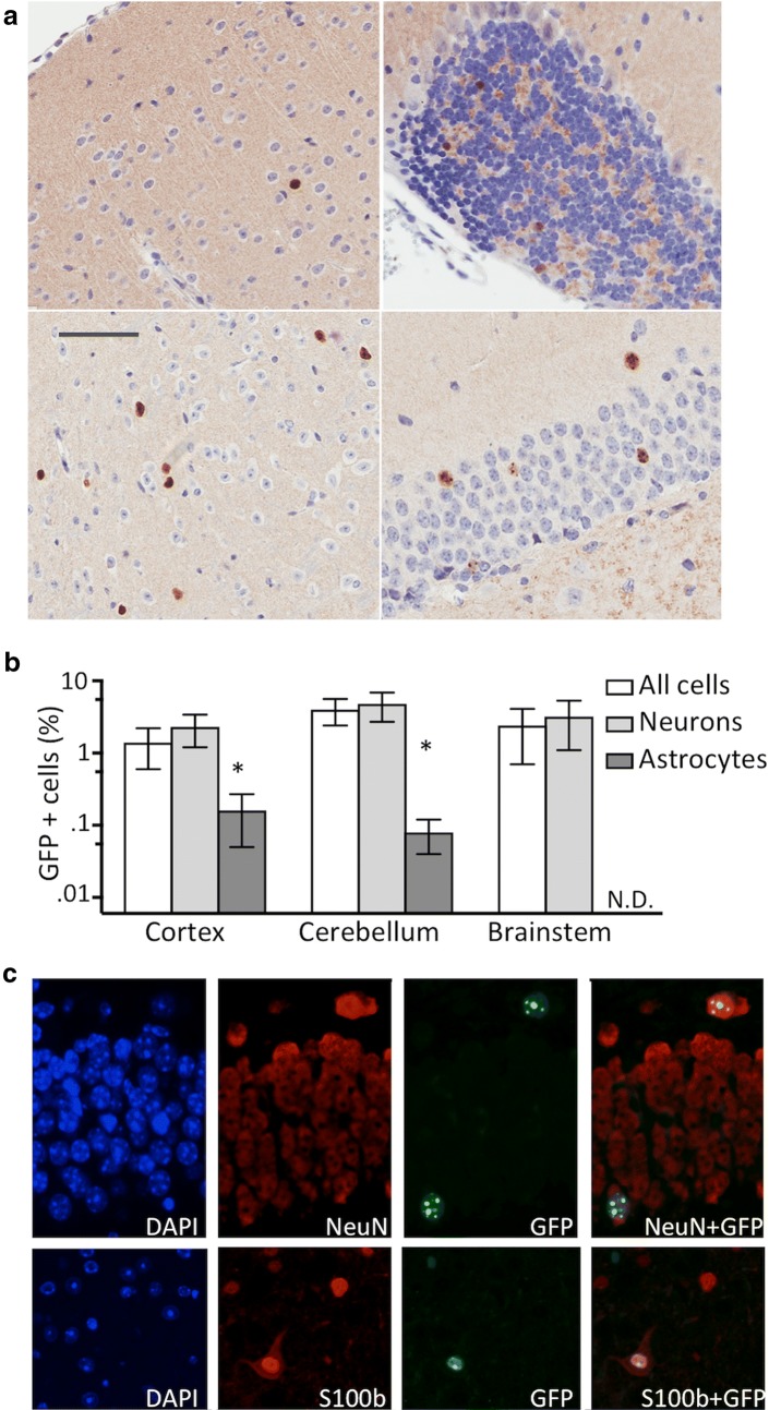
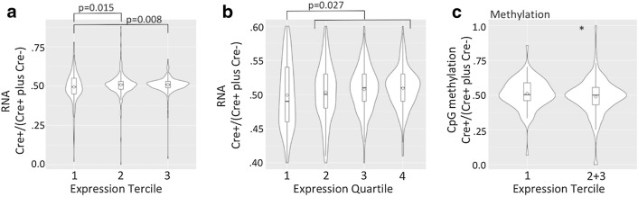
Results: By using an inactive X-chromosome-linked MeCP2-GFP reporter, which allowed us to enumerate reactivation events in the mouse brain even when they occur in very few cells, we found that deletion of Xist in the brain after establishment of X-chromosome inactivation leads to reactivation in 2-5% of neurons and in a smaller fraction of astrocytes. In contrast to global loss of both H3 lysine 27 trimethylation (H3K27m3) and histone H2A lysine 119 monoubiquitylation (H2AK119ub1) we observed upon Xist deletion, alterations in CpG methylation were subtle, and this was mirrored by only minor alterations in X-chromosome-wide gene expression levels, with highly expressed genes more prone to both derepression and demethylation compared to genes with low expression level.

Conclusion: Our results demonstrate that Xist plays a role in the maintenance of histone repressive marks, DNA methylation and transcriptional repression on the inactive X-chromosome, but that partial loss of X-dosage compensation in the absence of Xist in the brain is well tolerated.

Keywords: MeCP2; Noncoding RNA; Rett syndrome; X-chromosome inactivation; Xist.


Figures

Fig. 1
Fig. 1
a Mutation of Xist on the maternal X-chromosome leads to skewed inactivation of the MeCP2-GFP reporter gene on the paternal X-chromosome. b GFP immunohistochemistry with and without hematoxylin (+H and −H) in the brain of the MeCP2 Xistmut/MeCP2-GFP female demonstrates no GFP staining. The brain of the MeCP2/MeCP2-EGFP animal exhibits a bimodal nuclear staining for GFP reflective of random MeCP2-EGFP inactivation. c MeCP2-EGFP mRNA level in the brain from MeCP2 Xistmut/MeCP2-EGFP relative to MeCP2-EGFP/MeCP2 analyzed by RT-qPCR (n = 3 for each group, error bars indicate SD, ***p < 0.001, Student’s t test)
Fig. 2
Fig. 2
a Nestin-Cre deletes Xist2lox allele in the brain but not in the spleen of Xistmut/MeCP2-GFP-Xist2lox female animals. Ethidium bromide-stained gel of PCR products for Xist in the DNA extracted from the indicated tissues in animals with (Cre+) and without (Cre−) Nestin-Cre transgene. b Xist RNA level measured by RNA-seq in the RNA extracted from the Xistmut/MeCP2-GFP-Xist2lox females with (Cre+) and without (Cre−) Nestin-Cre transgene (n = 3). c Xistmut/ Xist2lox Nestin-Cre females that lack Xist in the brain were born at the expected Mendelian frequency (p = 0.36, Chi-square test). d Survival curves of females with Nestin-Cre transgene (nes +) and their littermates without (Cre−) the transgene (Cre+ n = 18, Cre− n = 17, N.S. p = 0.58, log-rank test). e Two-week sliding averages of weights in Xistmut/MeCP2-GFP Xist2lox females with (Cre+) and without (Cre−) Nestin-Cre transgene (Cre+ n = 14, Cre− n = 15, (**p < 0.01, Student’s t test, for all successive 2-week periods for the duration of the follow-up)
Fig. 3
Fig. 3
IF for a H3K27me3 and b H2AK119ub1 in the brain of animals without (control) and with Xist deletion (mutant). Punctate nuclear staining for both H3K27me3 and H2AK119ub1 is abolished by Xist deletion. c Percentage of cells in the brain with punctate staining for the indicated histone modification is severely reduced in animals lacking Xist (n = 3 for each group, error bar indicates SD, **p < 0.01, ***p < 0.001, Student’s t test)
Fig. 4
Fig. 4
a GFP IHC in different brain regions including cortex, hypocampus, brainstem and cerebellum (clockwise starting from left upper panel) of Nestin-Cre+ Xistmut/MeCP2-GFP Xist2lox animals lacking Xist in the brain. Bar indicates 80 µm. b Percentage of GFP-positive cells in the indicated brain regions in all cells, NeuN-positive neurons and S100b-positive astrocytes (n = 4, error bar indicates SD, neurons vs astrocytes, *p < 0.05, Student’s t test). c Reactivation of MeCP2-GFP reporter gene in neurons and astrocytes in brains from Nestin-Cre+ Xistmut/MeCP2-GFP Xist2lox animals. Double IF for GFP and NeuN (upper panel) and for GFP and S100b (lower panel) demonstrates GFP IF in both NeuN-positive neurons and S100b-positive astrocytes
Fig. 5
Fig. 5
Analysis of a global transcriptional and b DNA methylation changes on X-chromosome and autosomes in animals lacking Xist. a Left: Scatter plot of RNA expression levels for genes autosomes (black) and X-chromosome (red) (N = 3 for both mutant and control animals). The genes on X-chromosome exhibit an upward shift relative to autosomes. Middle: Cumulative expression plots of fold expression changes (mutant/control) for genes on autosomes (black) and X-chromosome (red). The genes on X-chromosome exhibit a rightward shift (p < 0.01, Wilcoxon rank-sum test). Right: Binned fold changes in expression for genes on autosomes (black) and X-chromosome (red). b Left: Scatter plot of DNA methylation abundance at CpG islands on autosomes (black) and X-chromosome (red) (N = 2 for both mutant and control animals). The CpG islands on X-chromosome exhibit a downward shift relative to CpG islands on autosomes. Middle: Cumulative plots of fold methylation abundance changes (mutant/control) for CpG islands on autosomes (black) and X-chromosome (red) (p < 0.01, Wilcoxon rank-sum test). Right: Binned fold changes in methylation abundance at CpG islands on autosomes (black) and X-chromosome (red)
Fig. 6
Fig. 6
Violin plots of the ratios of the RNA reads in Cre+/Cre+ plus Cre−) animals for X-linked genes that are divided into terciles (a) and quartiles (b) according to their expression level. Each tercile and quartile has 180 and 135 genes, respectively. All p values were calculated using Student’s t test. The average ratios for quartiles 1–4 are 0.494, 0.504, 0.508 and 0.510, respectively. The differences among adjacent quartiles are not significant. c Changes in CpG island methylation expressed as a ratio of methylation abundance in Cre+/(Cre+ plus Cre−) animals for genes grouped according to their expression level, as in (a), with the first tercile compared with an aggregate of the second and the third terciles (*p = 0.045, Student’s t test, 0.487 ± 0.133 vs 0.512 ± 0.109, N = 180 first tercile, N = 360 combined second and third terciles)

References

    1. Lyon MF. Gene action in the X-chromosome of the mouse (Mus musculus L.) Nature. 1961;190:372–373. doi: 10.1038/190372a0. - DOI - PubMed
    1. Maduro C, de Hoon B, Gribnau J. Fitting the puzzle pieces: the bigger picture of XCI. Trends Biochem Sci. 2016;41:138–147. doi: 10.1016/j.tibs.2015.12.003. - DOI - PubMed
    1. Disteche CM. Dosage compensation of the sex chromosomes. Annu Rev Genet. 2012;46:537–560. doi: 10.1146/annurev-genet-110711-155454. - DOI - PMC - PubMed
    1. Huynh KD, Lee JT. Inheritance of a pre-inactivated paternal X chromosome in early mouse embryos. Nature. 2003;426:857–862. doi: 10.1038/nature02222. - DOI - PubMed
    1. Okamoto I, Otte AP, Allis CD, Reinberg D, Heard E. Epigenetic dynamics of imprinted X inactivation during early mouse development. Science. 2004;303:644–649. doi: 10.1126/science.1092727. - DOI - PubMed

Publication types