. 2018 Sep 18;49(3):531-544.e6.
doi: 10.1016/j.immuni.2018.07.011. Epub 2018 Aug 28.

Lung γδ T Cells Mediate Protective Responses during Neonatal Influenza Infection that Are Associated with Type 2 Immunity

Affiliations

Lung γδ T Cells Mediate Protective Responses during Neonatal Influenza Infection that Are Associated with Type 2 Immunity

Xi-Zhi J Guo et al. Immunity. .

Abstract

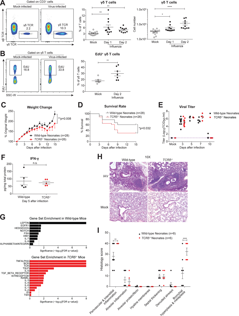
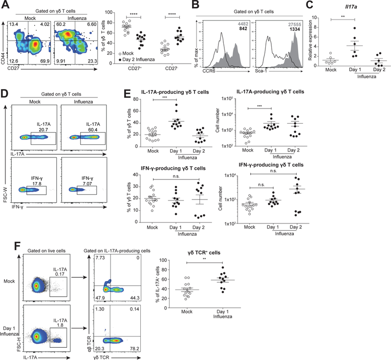
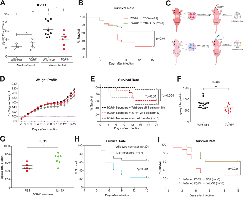
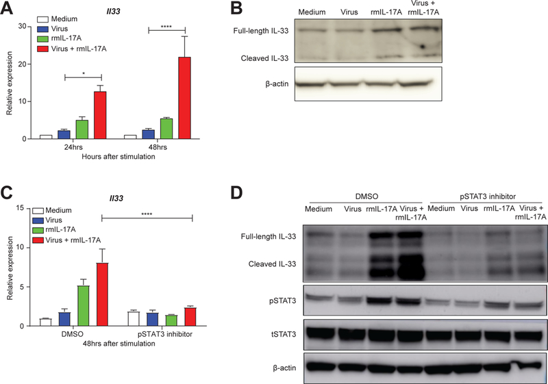
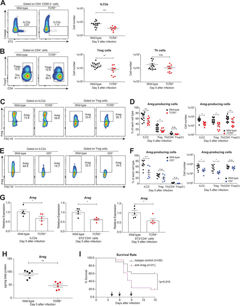
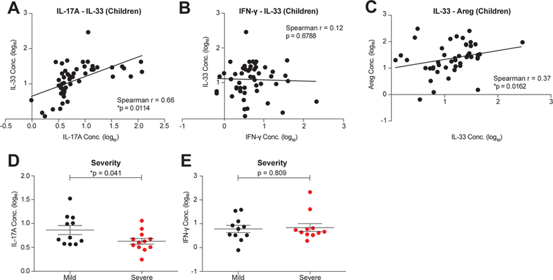
Compared to adults, infants suffer higher rates of hospitalization, severe clinical complications, and mortality due to influenza infection. We found that γδ T cells protected neonatal mice against mortality during influenza infection. γδ T cell deficiency did not alter viral clearance or interferon-γ production. Instead, neonatal influenza infection induced the accumulation of interleukin-17A (IL-17A)-producing γδ T cells, which was associated with IL-33 production by lung epithelial cells. Neonates lacking IL-17A-expressing γδ T cells or Il33 had higher mortality upon influenza infection. γδ T cells and IL-33 promoted lung infiltration of group 2 innate lymphoid cells and regulatory T cells, resulting in increased amphiregulin secretion and tissue repair. In influenza-infected children, IL-17A, IL-33, and amphiregulin expression were correlated, and increased IL-17A levels in nasal aspirates were associated with better clinical outcomes. Our results indicate that γδ T cells are required in influenza-infected neonates to initiate protective immunity and mediate lung homeostasis.

Keywords: IL-17A; IL-33; amphiregulin; children; neonatal influenza infection; type 2 immunity; γδ T cells.


Figures

Figure 1:
Figure 1:. γδ T cells protect neonatal mice against influenza infection via promotion of lung homeostasis and repair, independent of viral clearance.
A. Representative flow cytometric plots (left), and frequency and number (right) of γδ T cells in mock- (open circle, n=14) or virus-infected (solid) lungs of wild-type neonates at 1 (n=11) and 2 (n=10) days following intranasal influenza A/x31 virus infection. Data are combined from four independent experiments and presented as mean ± SEM.B. Representative flow cytometric plots (left) and summary frequency plot (right) of EdU+ γδ T cells in mock-infected (n=7) and influenza virus-infected (n=7) lungs of wild-type neonates 2 days after infection. Data are combined from three independent experiments and shown as mean ± SEM.C and D. Body weight profile (% of original weight) (C) and survival rate (D) of wild-type (black, n=28) and TCRδ−/− (red, n=26) neonates following influenza infection. Data are combined from sixteen independent experiments, and weight data are shown as mean ± SEM in change.E. Viral titer (Log10TCID50/ml) of wild-type (black) and TCRδ−/− (red) neonates assessed by plaque assay at days 0, 3, 5, 7 and 10 after influenza infection. Samples are pooled from at least two independent experiments for each time point and data are presented as mean ± SEM.F. Measurement of IFN-γ in the total lung homogenates by ELISA of wild-type (black, n=5) and TCRδ−/− (red, n=5) neonates at 7 days after influenza infection. Samples are pooled from three independent experiments, and data are presented as mean ± SEM.G. Gene Set Enrichment Analysis of whole-lung gene expression, ranked by significance (-Log10[FDR q-value]), from wild-type (black, n=3) and TCRδ−/− (red, n=3) neonates at 8 days after influenza infection.H. Representative images of H&E staining of influenza infected wild-type and TCRδ−/− lungs at 15 days after infection.I. Summary of histological analysis from influenza-infected wild-type (black, n=8) and TCRδ−/− (red, n=6) lungs at 15 days after infection. H-I. Data are combined from two independent experiments and shown as mean ± SEM.*p<0.05, **p<0.01, ****p<0.0001, n.s., not significant.
Figure 2
Figure 2. IL-17A-producing γδ T cells rapidly accumulate and respond to influenza infection in neonatal mice.
A. Representative flow cytometric plots (left, with CD27 and CD44 expression on x- and y-axis, respectively) and summary frequency plot (right) of γδ T cells from mock- (open, n=12) and influenza virus- (solid, n=11) infected mice at 2 days following infection. Data are combined from four independent experiments and presented as mean ± SEM.B. Representative flow cytometric histogram showing expression of CCR6 (left) and Sca-1 (right) gated on γδ T cells from mock- (open) and virus- (shaded) infected neonates (2 days after infection). Mean fluorescence intensity are shown in the upper right corners.C. Relative expression of Il17a by quantitative real-time PCR in sort-purified γδ T cells from mock (open, n=6) and virus-infected neonates at 1 (n=6) and 2 (n=6) days following infection. Samples are combined from two independent experiments and data are presented as mean ± SEM.D. Representative flow cytometric plots of IL-17A (top) and IFN-γ (bottom) expression in γδ T cells from mock- (left) and virus-infected (right) neonates at 1 day after infection.E. Scatter plot showing frequency and number of IL-17A- (top) and IFN-γ- (bottom) producing γδ T cells from (D). Samples from mock-infected (n=14) neonates were pooled from animals 1 (n=11) and 2 (n=10) days after mock infection.F. Representative flow cytometric plots (left) and summary frequency plots (right) of γδ TCR expression gated on total IL-17A-producing cells from mock- (n=14) and virus- (n=11) infected mice at 1 day following infection. (D-F) PMA/ionomycin was used for stimulation prior to intracellular staining. Data are combined from four independent experiments and presented as mean ± SEM. **p<0.01, ***p<0.001, ****p<0.0001, n.s., not significant.
Figure 3
Figure 3. IL-17A, predominantly secreted by γδ T cells, improves the survival of influenza-infected neonates by promoting IL-33 production.
A. Estimation of IL-17A in whole lung homogenates from mock- (open) or influenza virus-(solid)- infected wild-type (black) and TCRδ−/− (red) neonates by ELISA at 1 day after infection. Samples are pooled from at least two independent experiments and data are shown as mean ± SEM (Mann-Whitney test).B. Survival rate of influenza virus-infected TCRδ−/− neonates administered with low levels of recombinant mouse IL-17A (rmIL-17A, green, n=37, 100pg/mouse) or PBS control (red, n=19) at the time of infection. Data are combined from six independent trials, which individually showed the same trend, and data are presented as mean ± SEM.C. Schema outlining wild-type and Il17a−/− γδ T cell transfers to TCRδ−/− neonates and subsequent infection.D and E. Body weight profile (D) and survival rate (E) of influenza-infected TCRδ−/− neonates receiving wild-type (black, n=15) or Il17a−/− (red, n=13) γδ T cells intranasally or no cell transfer (grey, n=10). Data are combined from four independent trials, which individually showed the same trend. Weight profile data are presented as mean ± SEM.F. Protein levels of IL-33 assessed by ELISA in influenza virus-infected wild-type (black, n=15) and TCRδ−/− lungs (red, n=9) at 1 day following infection. Samples are pooled from three independent experiments, and data are shown as mean ± SEM.G. Protein levels of IL-33 detected by ELISA in the lungs of TCRδ−/− neonates that had been infected with influenza virus and simultaneously treated with either a low level of rmIL-17A (green, n=7, 100pg/mouse) or PBS control (red, n=6). Data were collected 1 day after infection/treatment. Samples are pooled from at least two independent experiments, and data are shown as mean ± SEM.H. Survival rate of influenza virus-infected wild-type (black, n=20) and Il33−/− neonates (blue, n=17) with intranasal influenza virus infection. Data are combined from ten independent experiments and shown as mean ± SEM in weight change.I. Survival rate of influenza virus-infected TCRδ−/− neonates administered with recombinant mouse IL-33 (rmIL-33, brown, n=18, 10ng/mouse) or PBS control (red, n=18) at the time of infection. Data are combined from six individual experiments.*p<0.05, **p<0.01, n.s., not significant.
Figure 4
Figure 4. IL-17A elevates IL-33 production in murine lung epithelial cells via STAT3 phosphorylation
A. Relative expression of Il33 mRNA, as assessed by quantitative real-time PCR, in mouse lung epithelial cells (LET1s) treated with medium (white), A/x31 influenza virus (blue, 3 MOI), rmIL-17A (green, 50ng/ml), or virus+rmIL-17A (red) at 24hrs and 48hrs after treatment. Data are shown as mean ± SEM and combined from three separate experiments that independently showed the same trend.B. Immunoblot assay of IL-33 protein in stimulated LET1s for the same conditions as in (A) at 48 hrs after stimulation.C. Quantitative real-time PCR analysis of Il33 mRNA in LET1 cells treated as in A and B with and without STAT3 phosphorylation inhibitor S3I-201 (100μM in 0.05% DMSO) at 48hrs after treatment. Data are shown as mean ± SEM and combined from two separate experiments that independently showed the same trend.D. Immunoblot analysis of IL-33 and phosphorylated STAT3 (pSTAT3) in the LET1 lysates treated as before (A, B, and C). Total STAT3 (tSTAT3) and β-actin are also shown. Immunoblots were repeated twice showing similar results.*p<0.05, ****p<0.0001
Figure 5.
Figure 5.. Deficiency in γδ T cells results in decreased accumulation and functionality of ILC2s and Treg cells.
A. Representative flow cytometric plots (left) and cell number (right) of ILC2s (CD4- CD90.2+ Lin- ST2+) in the lungs of infected wild-type (black, n=14) and TCRδ−/− (red, n=10) neonates at day 5 following infection.B. Representative flow cytometric plots (left) and cell number (right) of Treg cells (CD4+ Foxp3+) and Th cells (CD4+ Foxp3-) in the lungs of infected wild-type (black, n=14) and TCRδ−/− (red, n=10) neonates at day 5 following infection.C. Representative flow cytometric plots of Areg-producing cells gated on ILC2s and Treg cells in the lungs of infected wild-type (black, n=14) and TCRδ−/− (red, n=10) neonates at 5 days following infection.D. Frequency (left) and cell number (right) of Areg-producing ILC2s, Treg cells, and Th cells in the lungs of infected wild-type (black, n=14) and TCRδ−/− (red, n=10) neonates at 5 days after infection. (A-D) Data are combined from four independent experiments and shown as mean ± SEM.E. Representative flow cytometric plots of Areg-producing cells gated on ILC2s and Treg cells in the lungs of infected wild-type (black, n=8) and Il33−/− (blue, n=6) neonates at 5 days following infection.F. Frequency (left) and cell number (right) of Areg-producing ILC2s, Treg cells, and Th cells in the lungs of infected wild-type (black, n=8) and Il33−/− (blue, n=6) neonates at 5 days after infection. (E-F) Data are combined from three independent experiments and shown as mean ± SEM.G. Relative expression levels of Areg of ILC2 cells, ST2+CD4+ cells, and ST2-CD4+ cells sorted from wild-type (black, n=5) and TCRδ−/− (red, n=5) neonatal lungs at 5 days after infection measured by quantitative real-time PCR. Samples are pooled from two independent experiments, and data are presented as mean ± SEM.H. Protein levels of Areg in whole-lung lysate from infected wild-type (black, n=6) and TCRδ−/− (red, n=6) neonates at 5 days after infection. Data are combined from two independent experiments and shown as mean ± SEM.I. Survival rate of infected wild-type neonates that were injected intraperitoneally at days 4, 6, and 9 (shown by arrows) after influenza virus infection with Areg neutralizing antibody (purple, α-Areg antibody, 2ug/mouse/timepoint, n=21) or isotype control antibody (black, normal goat IgG control, 2ug/mouse/time point, n=20). Data are combined from three separate trials that individually showed the same trend.*p<0.05, **p<0.01, ***p<0.001, n.s., not significant.
Figure 6
Figure 6. The IL-17A/IL-33/Areg cascade in influenza-infected children is associated with robust disease outcome
A- C. Correlation between concentrations of human IL-17A and IL-33 (A), IFN-γ and IL-33 (B) and IL-33 and Areg protein levels (C) in the nasal aspirates of influenza-infected children (< 8 years old, n=51).D. Concentration of IL-17A at Day 0 after enrollment in the nasal aspirates of children with mild (black, n=11) and severe (red, n=12) influenza disease outcomes.E. Concentration of IFN-γ at Day 0 after enrollment in the nasal aspirates of children with mild (black, n=11) and severe (red, n=12) disease outcomes.Data are presented as mean ± SEM, and in each case cytokine values (pg/ml) were log10 transformed for visualization only. *p<0.05.

Similar articles

Cited by

References

    1. Adkins B, Leclerc C, and Marshall-Clarke S (2004). Neonatal adaptive immunity comes of age. Nat. Rev. Immunol. 4, 553–564. - PubMed
    1. Anders S, Pyl PT, and Huber W (2015). HTSeq--a Python framework to work with high-throughput sequencing data. Bioinformatics 31, 166–169. - PMC - PubMed
    1. Arpaia N, Green JA, Moltedo B, Arvey A, Hemmers S, Yuan S, Treuting PM, and Rudensky AY (2015). A Distinct Function of Regulatory T Cells in Tissue Protection. Cell 162, 1078–1089. - PMC - PubMed
    1. Bates D, Mächler M, Bolker B, and Walker S (2015). Fitting Linear Mixed-Effects Models Using lme4. Journal of Statistical Software, Articles 67, 1–48.
    1. Bhat N, Wright JG, Broder KR, Murray EL, Greenberg ME, Glover MJ, Likos AM, Posey DL, Klimov A, Lindstrom SE, et al. (2005). Influenza-Associated Deaths among Children in the United States, 2003–2004. N. Engl. J. Med. 353, 2559–2567. - PubMed

Publication types