Clinical Trial
. 2019 Jan;104(1):128-137.
doi: 10.3324/haematol.2017.186320. Epub 2018 Aug 31.

Clinical and molecular characteristics of MEF2D fusion-positive B-cell precursor acute lymphoblastic leukemia in childhood, including a novel translocation resulting in MEF2D-HNRNPH1 gene fusion

Affiliations
Clinical Trial

Clinical and molecular characteristics of MEF2D fusion-positive B-cell precursor acute lymphoblastic leukemia in childhood, including a novel translocation resulting in MEF2D-HNRNPH1 gene fusion

Kentaro Ohki et al. Haematologica. 2019 Jan.

Abstract

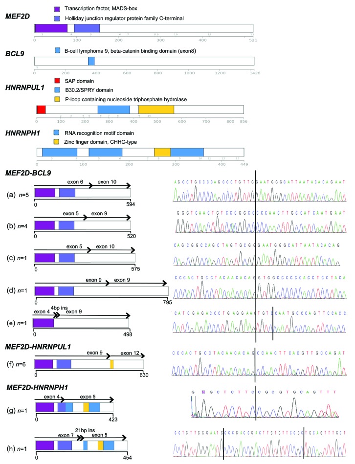
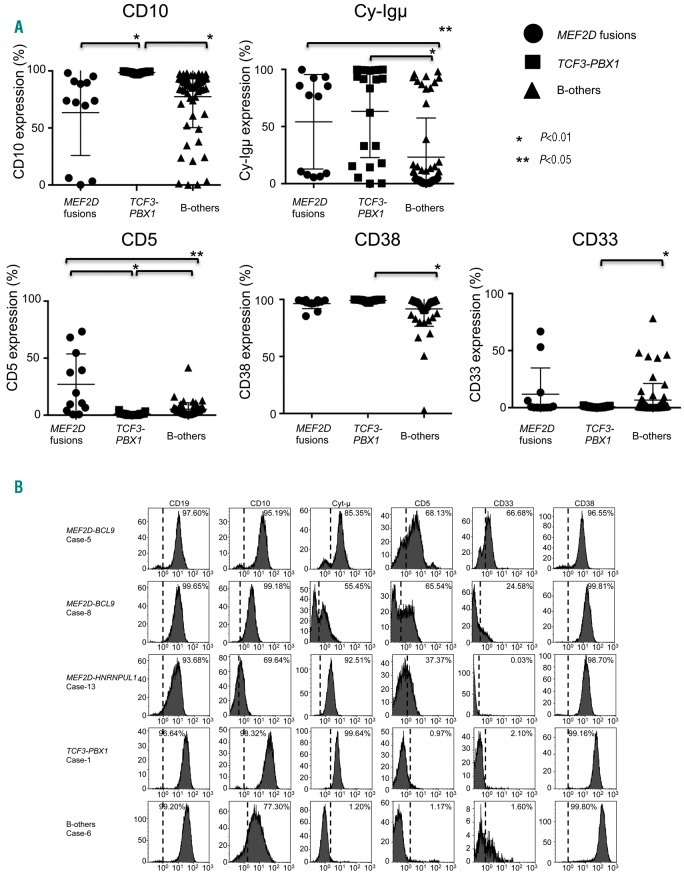
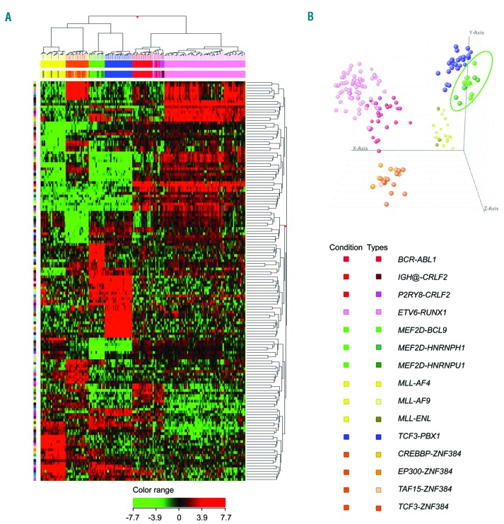
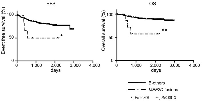
Fusion genes involving MEF2D have recently been identified in precursor B-cell acute lymphoblastic leukemia, mutually exclusive of the common risk stratifying genetic abnormalities, although their true incidence and associated clinical characteristics remain unknown. We identified 16 cases of acute lymphoblastic leukemia and 1 of lymphoma harboring MEF2D fusions, including MEF2D-BCL9 (n=10), MEF2D-HNRNPUL1 (n=6), and one novel MEF2D-HNRNPH1 fusion. The incidence of MEF2D fusions overall was 2.4% among consecutive precursor B-cell acute lymphoblastic leukemia patients enrolled onto a single clinical trial. They frequently showed a cytoplasmic μ chain-positive pre-B immunophenotype, and often expressed an aberrant CD5 antigen. Besides up- and down-regulation of HDAC9 and MEF2C, elevated GATA3 expression was also a characteristic feature of MEF2D fusion-positive patients. Mutations of PHF6, recurrent in T-cell acute lymphoblastic leukemia, also showed an unexpectedly high frequency (50%) in these patients. MEF2D fusion-positive patients were older (median age 9 years) with elevated WBC counts (median: 27,300/ml) at presentation and, as a result, were mostly classified as NCI high risk. Although they responded well to steroid treatment, MEF2D fusion-positive patients showed a significantly worse outcome, with 53.3% relapse and subsequent death. Stem cell transplantation was ineffective as salvage therapy. Interestingly, relapse was frequently associated with the presence of CDKN2A/CDKN2B gene deletions. Our observations indicate that MEF2D fusions comprise a distinct subgroup of precursor B-cell acute lymphoblastic leukemia with a characteristic immunophenotype and gene expression signature, associated with distinct clinical features.


Figures

Figure 1.
Figure 1.
Structures of the MEF2D fusions. Structures of fusion proteins and nucleotide sequences of (a) - (e) MEF2D-BCL9, (f) MEF2D-HNRNPUL1, (g) and (h) MEF2D-HNRNPH1. The red arrowhead shows the donor and acceptor site breakpoint. The number of patients in whom a particular fusion isoform was found is indicated on the right.
Figure 2.
Figure 2.
Immunophenotypic characteristics of B-ALL patients with MEF2D fusions. (A) The positivity (percentage) of CD10, cytoplasmic m, CD5, CD38, and CD33 of MEF2D fusion-positive, TCF3-PBX1-positive, and B-other patients are plotted as a scattergram. A detailed list of positivity for each immunophenotypic marker of the patients is presented in Online Supplementary Table S4. (B) Typical histograms of CD10, CD19, cytoplasmic m, aberrant CD5, CD33, and CD38 of MEF2D-BCL9, MEF2D-HNRNPUL1, TCF3-PBX1, and B-other patients are indicated with a positive rate (%). X-axis, fluorescence intensity; Y-axis, relative cell number.
Figure 3.
Figure 3.
Characteristics of gene expression profile in MEF2D-fusion-positive ALL. (A) Two-way hierarchical clustering and (B) principal component analysis (PCA) were performed on the microarray data, including B-ALLs with MEF2D fusion-positive and other types of genetic abnormalities, using the probe sets of differentially expressed genes between B-ALL with MEF2D fusions and other types of genetic abnormalities selected from filtered microarray probes (presented in Online Supplementary Tables S7 and S8). The results of clustering analysis are displayed using a heat map as a dendrogram.
Figure 4.
Figure 4.
Outcomes of patients with MEF2D fusions. (A) Kaplan-Meier estimates of event-free survival (EFS) of patients with MEF2D fusions, and B-other, log-rank P=0.306). (B) Overall survival (OS) for the same as above (log-rank P=0.0013).

Similar articles

Cited by

References

    1. Hunger SP, Mullighan CG. Acute lymphoblastic leukemia in children. N Engl J Med. 2015;373(16):1541–1552. - PubMed
    1. Schwab C, Harrison CJ. Advances in B-cell precursor acute lymphoblastic leukemia genonics. HemaSphere. 2018;2(4):e53 - PMC - PubMed
    1. Roberts KG, Morin RD, Zhang J, et al. Genetic alterations activating kinase and cytokine receptor signaling in high-risk acute lymphoblastic leukemia. Cancer Cell. 2012;22(2):153–166. - PMC - PubMed
    1. Roberts KG, Li Y, Payne-Turner D, et al. Targetable kinase-activating lesions in Ph-like acute lymphoblastic leukemia. N Engl J Med. 2014;371(11):1005–1015. - PMC - PubMed
    1. Gocho Y, Kiyokawa N, Ichikawa H, et al. A novel recurrent EP300-ZNF384 gene fusion in B-cell precursor acute lymphoblastic leukemia. Leukemia. 2015;29(12):2445–2448. - PubMed

Publication types

MeSH terms

Substances