. 2018 Oct 26;293(43):16635-16646.
doi: 10.1074/jbc.RA118.002425. Epub 2018 Aug 31.

Differential biochemical properties of three canonical Dps proteins from the cyanobacterium Nostoc punctiforme suggest distinct cellular functions

Affiliations

Differential biochemical properties of three canonical Dps proteins from the cyanobacterium Nostoc punctiforme suggest distinct cellular functions

Christoph Howe et al. J Biol Chem. .

Abstract

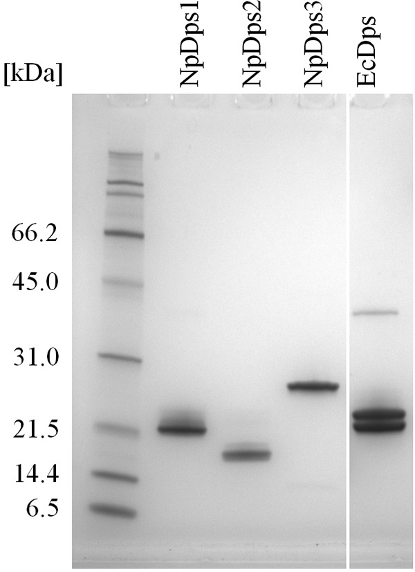
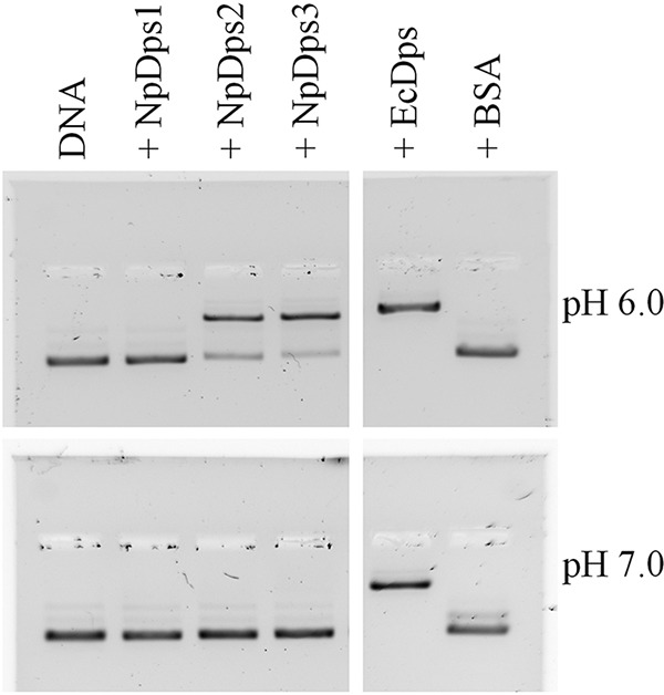
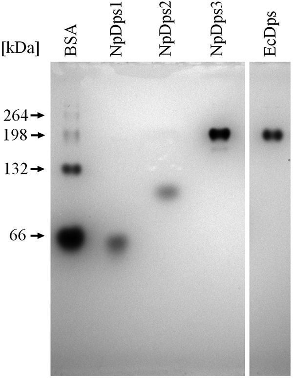
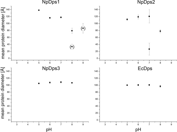
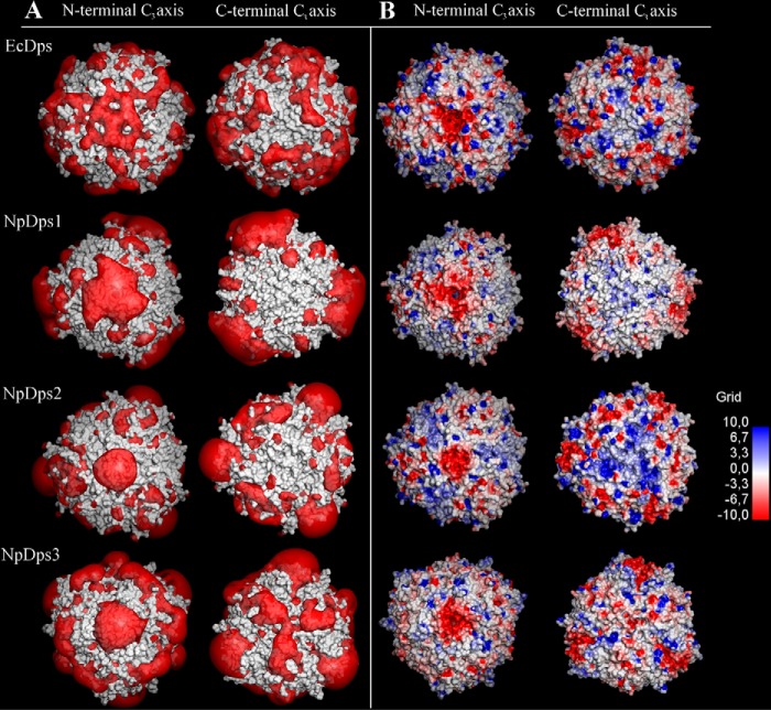
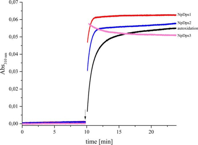
DNA-binding proteins from starved cells (Dps, EC: 1.16.3.1) have a variety of different biochemical activities such as DNA-binding, iron sequestration, and H2O2 detoxification. Most bacteria commonly feature one or two Dps enzymes, whereas the cyanobacterium Nostoc punctiforme displays an unusually high number of five Dps proteins (NpDps1-5). Our previous studies have indicated physiological differences, as well as cell-specific expression, among these five proteins. Three of the five NpDps proteins, NpDps1, -2, and -3, were classified as canonical Dps proteins. To further investigate their properties and possible importance for physiological function, here we characterized and compared them in vitro Nondenaturing PAGE, gel filtration, and dynamic light-scattering experiments disclosed that the three NpDps proteins exist as multimeric protein species in the bacterial cell. We also demonstrate Dps-mediated iron oxidation catalysis in the presence of H2O2 However, no iron oxidation with O2 as the electron acceptor was detected under our experimental conditions. In modeled structures of NpDps1, -2, and -3, protein channels were identified that could serve as the entrance for ferrous iron into the dodecameric structures. Furthermore, we could demonstrate pH-dependent DNA-binding properties for NpDps2 and -3. This study adds critical insights into the functions and stabilities of the three canonical Dps proteins from N. punctiforme and suggests that each of the Dps proteins within this bacterium has a specific biochemical property and function.

Keywords: DNA binding protein; DNA-binding proteins from starved cells; bacterial metabolism; cyanobacteria; ferritin; iron; metal homeostasis; multifunctional protein; oxidative stress; reactive oxygen species (ROS).


Conflict of interest statement

The authors declare that they have no conflicts of interest with the contents of this article

Figures

Figure 1.
Figure 1.
SDS-PAGE analysis on three recombinant Dps proteins from N. punctiforme and E. coli Dps (EcDps). 1 μg of each of the purified proteins was loaded on an SDS-PAGE gel. Expected molecular masses of the Dps monomers (including the alanine-alanine linker and the Strep(II)-tag sequence) were: 21.2 kDa for NpDps1, 19.5 kDa for NpDps2, 22.2 kDa for NpDps3, and 19.9 kDa for EcDps. SDS-PAGE broad range molecular weight standards (Bio-Rad) were used as molecular mass markers. The gel was stained with colloidal Coomassie.
Figure 2.
Figure 2.
EMSA to analyze the DNA-binding properties of the NpDps proteins under different pH conditions. At pH 6.0 and 7.0, 125 ng of plasmid DNA (pSB1A3 vector) was incubated with 1 μg of each Dps protein, and separated on an agarose gel (1%). E. coli Dps (EcDps) and BSA served as a positive and negative control, respectively. Plasmid DNA is shown in lane DNA. Thiazole orange staining was performed after gel electrophoresis for DNA detection. The gel documentations are shown in inverted colors.
Figure 3.
Figure 3.
Multimerization of NpDps1, NpDps2, and NpDps3 and E. coli Dps (EcDps) analyzed by nondenaturing PAGE. The theoretical molecular masses of putative dodecamers were 253 kDa for NpDps1, 233 kDa for NpDps2, 265 kDa for NpDps3, and 236 kDa for EcDps. Additionally, the different native forms of BSA were used to estimate the approximate molecular masses. The pH of the gel was 8.5 and the pH of the running buffer was 8.0. The gel was stained with colloidal Coomassie.
Figure 4.
Figure 4.
DLS on NpDps1, NpDps2, and NpDps3 at different pH conditions. The hydrodynamic diameters of all protein species are displayed as a mean of the average hydrodynamic diameter from each experimental series in Å at pH 3.0, 5.0, 6.0, 7.0, 8.0, and pH 9.0. Particles with a lower peak occurrence than 1% (number related) are not displayed, whereas particles with occurrence between 1 and 10% are shown in parentheses. The E. coli Dps (EcDps) served as a control. Mean ± S.E. particle diameters are given as error bars.
Figure 5.
Figure 5.
Calculated electrostatic potential around the N- and C-terminal C3-axes in E. coli Dps (EcDps) and the model structures of NpDps1, NpDps2, and NpDps3. A, electrostatic potential isosurfaces (red; −1.0 kT/e) of the Dps proteins, superimposed onto the van der Waals surface of the proteins (gray). B, the van der Waals surfaces of the Dps proteins colored according to the calculated electrostatic potentials from electronegative (red) to electropositive (blue) by a continuous color gradient. The N-terminal C3-axes indicates the ferritin-like pore and the C-terminal C3-axes indicates the Dps-type pore.
Figure 6.
Figure 6.
Fe2+ oxidation kinetics in the presence of NpDps1, NpDps2, and NpDps3. Absorbance measured at 310 nm corresponds to the formation of the ferric iron in the presence of NpDps1 (red line), NpDps2 (blue line), NpDps3 (pink line), and for the no protein control (black line). 0.5 μm of each NpDps protein was mixed with 24 μm Fe2+ (48 iron ions per Dps dodecamer), at time 0 min and the absorbance was analyzed for 10 min, before addition of 16 μm H2O2 as indicated by the arrow. Reactions were performed in 5 mm succinate buffer, pH 6.0, 50 mm NaCl, at room temperature and under aerobic conditions.

References

    1. Imlay J. A. (2006) Iron-sulphur clusters and the problem with oxygen. Mol. Microbiol. 59, 1073–1082 10.1111/j.1365-2958.2006.05028.x - DOI - PubMed
    1. Andrews S. C., Robinson A. K., and Rodríguez-Quiñones F. (2003) Bacterial iron homeostasis. FEMS Microbiol. Rev. 27, 215–237 10.1016/S0168-6445(03)00055-X - DOI - PubMed
    1. Zhao G., Ceci P., Ilari A., Giangiacomo L., Laue T. M., Chiancone E., and Chasteen N. D. (2002) Iron and hydrogen peroxide detoxification properties of DNA-binding protein from starved cells. A “ferritin-like” DNA-binding protein of Escherichia coli. J. Biol. Chem. 277, 27689–27696 10.1074/.M202094200 - DOI - PubMed
    1. Calhoun L. N., and Kwon Y. M. (2011) Structure, function and regulation of the DNA-binding protein Dps and its role in acid and oxidative stress resistance in Escherichia coli: a review. J. Appl. Microbiol. 110, 375–386 - PubMed
    1. Ceci P., Cellai S., Falvo E., Rivetti C., Rossi G. L., and Chiancone E. (2004) DNA condensation and self-aggregation of Escherichia coli Dps are coupled phenomena related to the properties of the N-terminus. Nucleic Acids Res. 32, 5935–5944 10.1093/nar/gkh915 - DOI - PMC - PubMed

Publication types

Associated data