. 2018 Oct 15;201(8):2303-2314.
doi: 10.4049/jimmunol.1800732. Epub 2018 Aug 31.

Hepatitis B Virus Surface Antigen Enhances the Sensitivity of Hepatocytes to Fas-Mediated Apoptosis via Suppression of AKT Phosphorylation

Affiliations

Hepatitis B Virus Surface Antigen Enhances the Sensitivity of Hepatocytes to Fas-Mediated Apoptosis via Suppression of AKT Phosphorylation

Zhen-Tang Jing et al. J Immunol. .

Erratum in

Abstract

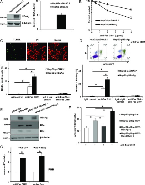
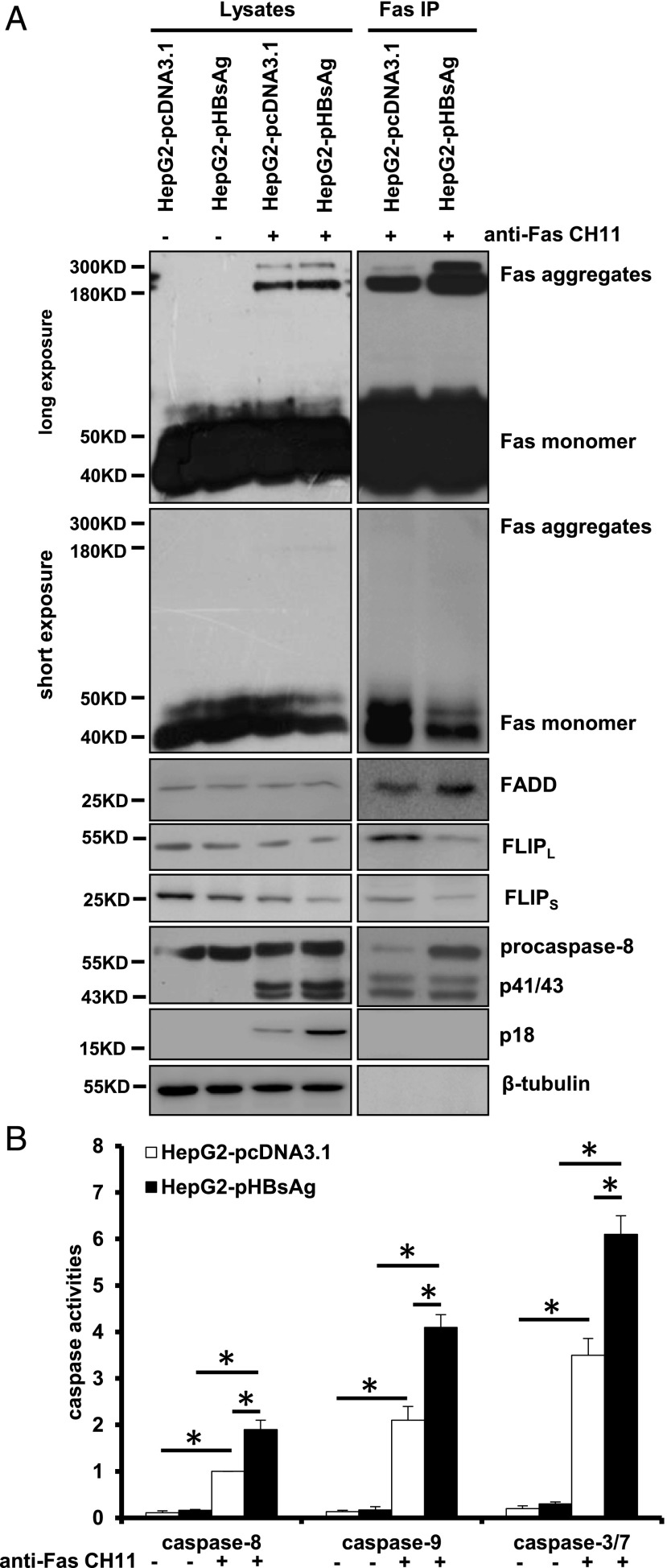
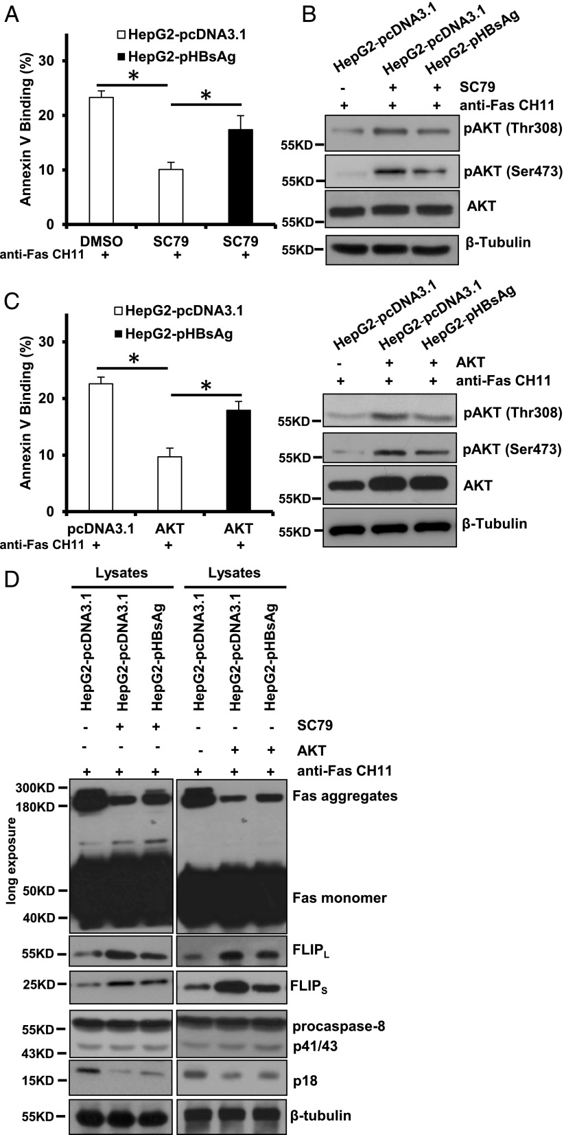
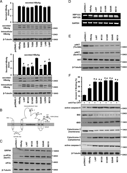
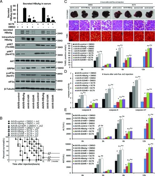
The Fas receptor/ligand system plays a prominent role in hepatic apoptosis and hepatocyte death. Although hepatitis B virus (HBV) surface Ag (HBsAg) is the most abundant HBV protein in the liver and peripheral blood of patients with chronic HBV infection, its role in Fas-mediated hepatocyte apoptosis has not been disclosed. In this study, we report that HBsAg sensitizes HepG2 cells to agonistic anti-Fas Ab CH11-induced apoptosis through increasing the formation of SDS-stable Fas aggregation and procaspase-8 cleavage but decreasing both the expression of cellular FLIPL/S and the recruitment of FLIPL/S at the death-inducing signaling complex (DISC). Notably, HBsAg increased endoplasmic reticulum stress and consequently reduced AKT phosphorylation by deactivation of phosphoinositide-dependent kinase-1 (PDPK1) and mechanistic target of rapamycin complex 2 (mTORC2), leading to enhancement of Fas-mediated apoptosis. In a mouse model, expression of HBsAg in mice injected with recombinant adenovirus-associated virus 8 aggravated Jo2-induced acute liver failure, which could be effectively attenuated by the AKT activator SC79. Based on these results, it is concluded that HBsAg predisposes hepatocytes to Fas-mediated apoptosis and mice to acute liver failure via suppression of AKT prosurviving activity, suggesting that interventions directed at enhancing the activation or functional activity of AKT may be of therapeutic value in Fas-mediated progressive liver cell injury and liver diseases.


Figures

FIGURE 1.
FIGURE 1.
Expression of HBsAg renders hepatoma cells and PHH hypersensitive to Fas-mediated apoptosis. (A) Forced expression of HBsAg in HepG2 cells was confirmed by Western blot analysis and Architect assay. (B) In vitro cytotoxicity of anti-Fas CH11 to HepG2-pcDNA and HepG2-pHBsAg cells. The cells were treated with increasing concentrations of anti-Fas CH11 in the presence of 0.5 μg/ml ActD for 24 h, and a CCK-8 assay was used to quantify cell viability. Values are mean ± SD; n = 5. (C) Quantification of apoptotic cell proportions by TUNEL staining. Cells were treated with 1 μg/ml anti-Fas CH11 in the presence of ActD for 4 h with or without pretreatment with antagonistic anti-Fas ZB4. Data are expressed as percentage of the number of TUNEL-positive cells to the total number of HepG2-pcDNA3.1 or HepG2-pHBsAg cells counted. Original magnification ×200. Values are mean ± SD; n = 5. *p < 0.05. (D) Quantification of apoptotic cell fractions by annexin V/propidium iodide (PI) staining. Cells received the same treatments as indicated in (C) and were subjected to flow cytometric analysis. Values are mean ± SD; n = 3. *p < 0.05. (E) Expression of HBsAg, HBc, and HBx in HepG2-pRep HBV, HepG2-pRep HBV HBsAg(−), and HepG2-pRep HBV HBc and HBx(−) cells was assessed by Western blot analysis. (F) Flow cytometric analysis of the fraction of apoptotic cells 4 h after the indicated cells were treated with 1 μg/ml CH11 in the presence of ActD. Values are mean ± SD; n = 3. *p < 0.05. (G) Measurements of caspase-3/7 enzymatic activity of Ad-GFP– or Ad-HBsAg–infected PHH after CH11 and FasL stimulation. Values are mean ± SD; n = 3. *p < 0.05.
FIGURE 2.
FIGURE 2.
HBsAg facilitates procaspase-8 cleavage via enhancing the formation of SDS-stable Fas aggregation and reducing the recruitment of FLIPL/S at the DISC. (A) Analysis of the DISC components from the immunoprecipitated complexes with anti-Fas Ab. (B) Measurements of caspase-3/7, -8, and -9 enzymatic activity expressed as the fold change relative to that in the HepG2-pcDNA3.1 control cells. Values are mean ± SD; n = 3. *p < 0.05.
FIGURE 3.
FIGURE 3.
HBsAg restored Fas-mediated apoptosis initially attenuated by enhanced AKT phosphorylation. (A) Quantification of apoptotic cell fractions by annexin V/propidium iodide staining. DMSO- or SC79-pretreated HepG2-pcDNA3.1 and HepG2-pHBsAg cells were exposed to 1 μg/ml agonistic Fas CH11 for 6 h and subjected to flow cytometric analysis. Values are mean ± SD; n = 3. *p < 0.05. (B) HBsAg abrogated SC79-induced Akt activation in HepG2 cells. (C) Quantification of apoptotic cell fractions by annexin V/PI staining in the AKT-overexpressed cells 6 h after treatment with 1 μg/ml agonistic Fas CH11. Values are mean ± SD; n = 3. *p < 0.05. Western blot analysis shows that HBsAg abrogated AKT activation by ectopic expression of AKT in HepG2 cells. (D) Analysis of the DISC components from the lysates stimulated with anti-Fas CH11.
FIGURE 4.
FIGURE 4.
HBsAg downregulates AKT phosphorylation via deactivation of PDPK1 and mTORC2. (A) Representative Western blot showing the effect of HBsAg on expression of total and phosphorylated forms of key molecules in the AKT signaling pathway in the HepG2-pcDNA3.1 and HepG2-pHBsAg cells. (B) Representative Western blot showing the effect of 4-PBA on expression of total and phosphorylated forms of key molecules in the AKT signaling pathway in the HepG2-pHBsAg cells. (C) Quantification of apoptotic cell fractions by annexin V/propidium iodide staining in the 4-PBA–pretreated HepG2-pcDNA3.1 and HepG2-pHBsAg cells 6 h after treatment with 1 μg/ml agonistic Fas CH11. Values are mean ± SD; n = 3. *p < 0.05. (D and E) Interaction between HBsAg and AKT, PDPK1, mTOR, and PTEN as determined by coimmunoprecipitation assay. FLAG-tagged HBsAg was immunoprecipitated with anti-FLAG Ab, and AKT, PDPK1, mTOR, or PTEN was detected by Western blotting with specific Abs. Reciprocal coimmunoprecipitation was carried out using AKT, PDPK1, mTOR, and PTEN Abs, and HBsAg was detected by Western blotting with anti-FLAG Ab. Normal IgG served as a control. (F) The effect of 4-PBA on interaction between mTOR and pmTOR (Ser2481), Rictor, AKT, and pAKT (Ser473) in HepG2-pcDNA3.1 and HepG2-pHBsAg cells as determined by coimmunoprecipitation assay. mTOR was immunoprecipitated with the specific Ab, and pmTOR (Ser2481), Rictor, AKT, or pAKT (Ser473) was detected by Western blotting with specific Abs.
FIGURE 5.
FIGURE 5.
HBsAg intracellular retention as a result of secretion deficiency enhances its proapoptotic activity by induction of ER stress. (A) Expression of secreted HBsAg from HepG2 cells stably transfected with wild-type and various mutated HBsAg was assessed by Western blot analysis and Architect assay. (B) Schematic illustration of HBsAg mutations sites responsible for intracellular retention of HBsAg and its proapoptotic activity. (C) Representative Western blot showing the expression of total and phosphorylated forms of key molecules involved in ER stress in HepG2 cells stably transfected with wild-type and mutated forms of HBsAg. (D) Semiquantitative RT-PCR showing the expression of XBP1(U) and XBP1(S) in HepG2 cells stably transfected with wild-type and mutated forms of HBsAg. (E) Representative Western blot showing the expression of total and phosphorylated forms of AKT in HepG2 cells stably transfected with wild-type and mutated forms of HBsAg. (F) Quantification of apoptotic cell fractions by annexin V/propidium iodide staining (upper panel) and Western blot analysis of expression of apoptosis-related mediators in HepG2 cells stably transfected with wild-type and mutated forms of HBsAg. Values are mean ± SD. n = 3. #p < 0.05, *p < 0.05.
FIGURE 6.
FIGURE 6.
Expression of HBsAg in mice renders them susceptible to ALF, and this effect is attenuated by administration of SC79. (A) SC79 treatment led to AKT hyperactivation in the livers of mice expressing wild-type and variant HBsAg by the AAV8 system. Protein extracts collected from the liver of untreated and SC79-treated mice were resolved on SDS-PAGE and immunoblotted with indicated Abs. SC79 was applied via i.p. injection at a concentration of 10 mg/kg body weight. (B) The protective effects of SC79 on an acute and lethal apoptotic hepatic injury induced by an anti-Fas agonist, Jo2 Ab. SC79-pretreated mice markedly extended the survival time from 6 to 13 h for AAV8-S204R, 6 to 14 h for AAV8-G145R, 9 to 15 h for AAV8-HBsAg, and 12 to 15 h for AAV8-control (all p < 0.01, n = 10 for all groups). (C) Macroscopic appearance of representative liver samples with H&E staining, and TUNEL staining 6 h after Jo2 treatment of the different groups as indicated. The histogram summarizes the average percentage of apoptotic cells in the livers of the indicated groups at various time points after Jo2 treatment. Original magnification ×200. (D) Caspase-8, -9, and -3/7 activity assay and (E) serum ALT and AST analyses, showing that significantly less severity with regard to apoptosis, caspase activities, and serum aminotransferase levels was observed in SC79-pretreated mice. Values are mean ± SD. n = 3. #p < 0.05, *p < 0.05.

References

    1. Nagata S. 1997. Apoptosis by death factor. Cell 88: 355–365. - PubMed
    1. Kondo T., Suda T., Fukuyama H., Adachi M., Nagata S. 1997. Essential roles of the Fas ligand in the development of hepatitis. Nat. Med. 3: 409–413. - PubMed
    1. Mochizuki K., Hayashi N., Hiramatsu N., Katayama K., Kawanishi Y., Kasahara A., Fusamoto H., Kamada T. 1996. Fas antigen expression in liver tissues of patients with chronic hepatitis B. J. Hepatol. 24: 1–7. - PubMed
    1. Lapinski T. W., Kowalczuk O., Prokopowicz D., Chyczewski L. 2004. Serum concentration of sFas and sFasL in healthy HBsAg carriers, chronic viral hepatitis B and C patients. World J. Gastroenterol. 10: 3650–3653. - PMC - PubMed
    1. Luo K. X., Zhu Y. F., Zhang L. X., He H. T., Wang X. S., Zhang L. 1997. In situ investigation of Fas/FasL expression in chronic hepatitis B infection and related liver diseases. J. Viral Hepat. 4: 303–307. - PubMed

Publication types

MeSH terms