

doi: 10.1038/s41592-018-0106-z.
Epub 2018 Aug 31.
CDeep3M-Plug-and-Play cloud-based deep learning for image segmentation
Affiliations
- PMID: 30171236
- PMCID: PMC6548193
- DOI: 10.1038/s41592-018-0106-z
Item in Clipboard
CDeep3M-Plug-and-Play cloud-based deep learning for image segmentation
Nat Methods.
2018 Sep.
Abstract
As biomedical imaging datasets expand, deep neural networks are considered vital for image processing, yet community access is still limited by setting up complex computational environments and availability of high-performance computing resources. We address these bottlenecks with CDeep3M, a ready-to-use image segmentation solution employing a cloud-based deep convolutional neural network. We benchmark CDeep3M on large and complex two-dimensional and three-dimensional imaging datasets from light, X-ray, and electron microscopy.
Conflict of interest statement
Competing interests
The authors declare no competing interests.
Figures
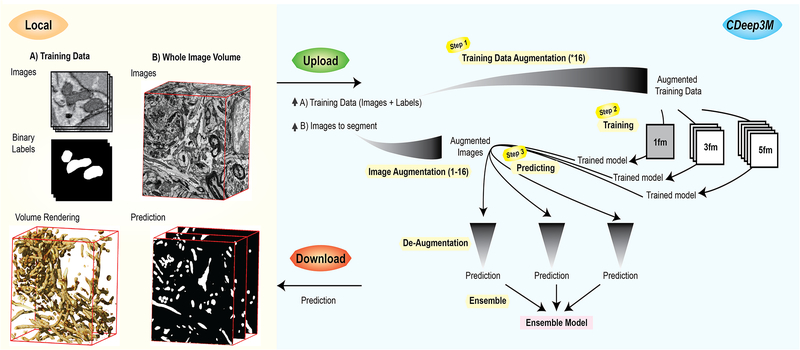
In Steps 1-2 a new trained model is generated, based on training images and labels. For 3D segmentation tasks CDeep3M trains three different models seeing 1 frame (1fm), seeing 3 frames (3fm) and seeing 5 frames (5fm) that are applied in Step 3 to provide three predictions. Those are merged into a single ensemble model at the post-processing step (Step 3).
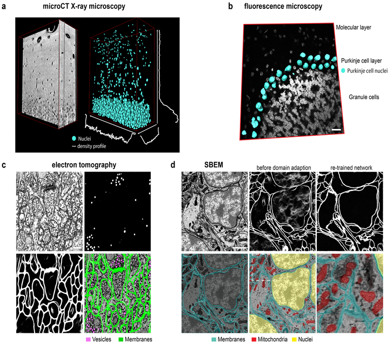
(a) Segmentation of nuclei in XRM volume of a 50μm mouse brain slice containing the hippocampal DG area used for cell counting and establishing a cell density profile across x-y-z. (b) Segmentation of cell type specific DNA profile allows identification of Purkinje cells. Overlay of 3D surface mesh of nuclei on light microscopic image of DAPI-stained mouse cerebellar brain section. Scale bar: 20μm. (c) Segmentation of vesicles and membranes on multi-tilt electron tomography of high-pressure frozen mouse brain section. Scale bar: 200nm. (d) Upper row: SBEM micrograph (left) Scale bar: 1μm, segmentation using pre-trained model before (middle) and after domain adaption (right). Lower row: segmentation of membranes, mitochondria and nuclei overlaid on SBEM data
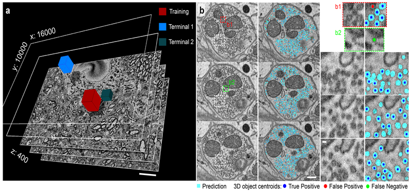
(a) SBEM volume acquired at 2.4nm × 24nm voxelsize (16000×10000×400 voxel). Performance tests were done on two terminals (Terminal 1: 3183 vesicles and Terminal 2: 1000 vesicles) comparing to several independent human counts. Scale: 5μm (b) Three consecutive sections of Terminal 1 in overview (left panels, Scale: 200nm) alongside the CDeep3M predictions and zoomed in (right panels, Scale: 40nm) show comparisons to human counts. Centroids of 3D objects occurring across sections are marked on most prevalent plane.
References
-
- Briggman KL, Helmstaedter M & Denk W Wiring specificity in the direction-selectivity circuit of the retina. Nature 471, 183–190 (2011). - PubMed
-
- Çiçek Ö, Abdulkadir A, Lienkamp SS, Brox T & Ronneberger O 3D U-net: Learning dense volumetric segmentation from sparse annotation. Lect. Notes Comput. Sci. (including Subser. Lect. Notes Artif. Intell. Lect. Notes Bioinformatics) 9901 LNCS, 424–432 (2016).
-
- Quan TM, Hildebrand DGC & Jeong W-K FusionNet: A deep fully residual convolutional neural network for image segmentation in connectomics. (2016).
Methods-only References
-
- Deerinck T et al. Enhancing Serial Block-Face Scanning Electron Microscopy to Enable High Resolution 3-D Nanohistology of Cells and Tissues. Microsc. Microanal 16, 1138–1139 (2010).
Publication types
MeSH terms
Grants and funding
LinkOut - more resources
Full Text Sources
Other Literature Sources
