. 2018 Sep;15(9):669-676.
doi: 10.1038/s41592-018-0085-0. Epub 2018 Aug 31.

Precision and accuracy of single-molecule FRET measurements-a multi-laboratory benchmark study

Björn Hellenkamp  1   2 Sonja Schmid  1   3 Olga Doroshenko  4 Oleg Opanasyuk  4 Ralf Kühnemuth  4 Soheila Rezaei Adariani  5 Benjamin Ambrose  6 Mikayel Aznauryan  7 Anders Barth  8 Victoria Birkedal  7 Mark E Bowen  9 Hongtao Chen  10 Thorben Cordes  11   12 Tobias Eilert  13 Carel Fijen  14 Christian Gebhardt  12 Markus Götz  1 Giorgos Gouridis  11   12 Enrico Gratton  10 Taekjip Ha  15 Pengyu Hao  16 Christian A Hanke  4 Andreas Hartmann  17 Jelle Hendrix  18   19 Lasse L Hildebrandt  7 Verena Hirschfeld  20 Johannes Hohlbein  14   21 Boyang Hua  15 Christian G Hübner  20 Eleni Kallis  13 Achillefs N Kapanidis  22 Jae-Yeol Kim  23 Georg Krainer  17   24 Don C Lamb  8 Nam Ki Lee  23 Edward A Lemke  25   26   27 Brié Levesque  9 Marcia Levitus  28 James J McCann  9 Nikolaus Naredi-Rainer  8 Daniel Nettels  29 Thuy Ngo  15 Ruoyi Qiu  16 Nicole C Robb  22 Carlheinz Röcker  13 Hugo Sanabria  5 Michael Schlierf  17 Tim Schröder  30 Benjamin Schuler  29 Henning Seidel  20 Lisa Streit  13 Johann Thurn  1 Philip Tinnefeld  30   31 Swati Tyagi  27 Niels Vandenberk  18 Andrés Manuel Vera  30 Keith R Weninger  16 Bettina Wünsch  31 Inna S Yanez-Orozco  5 Jens Michaelis  32 Claus A M Seidel  33 Timothy D Craggs  34   35 Thorsten Hugel  36   37
Affiliations

Precision and accuracy of single-molecule FRET measurements-a multi-laboratory benchmark study

Björn Hellenkamp et al. Nat Methods. 2018 Sep.

Erratum in

  • Publisher Correction: Precision and accuracy of single-molecule FRET measurements-a multi-laboratory benchmark study.
    Hellenkamp B, Schmid S, Doroshenko O, Opanasyuk O, Kühnemuth R, Adariani SR, Ambrose B, Aznauryan M, Barth A, Birkedal V, Bowen ME, Chen H, Cordes T, Eilert T, Fijen C, Gebhardt C, Götz M, Gouridis G, Gratton E, Ha T, Hao P, Hanke CA, Hartmann A, Hendrix J, Hildebrandt LL, Hirschfeld V, Hohlbein J, Hua B, Hübner CG, Kallis E, Kapanidis AN, Kim JY, Krainer G, Lamb DC, Lee NK, Lemke EA, Levesque B, Levitus M, McCann JJ, Naredi-Rainer N, Nettels D, Ngo T, Qiu R, Robb NC, Röcker C, Sanabria H, Schlierf M, Schröder T, Schuler B, Seidel H, Streit L, Thurn J, Tinnefeld P, Tyagi S, Vandenberk N, Vera AM, Weninger KR, Wünsch B, Yanez-Orozco IS, Michaelis J, Seidel CAM, Craggs TD, Hugel T. Hellenkamp B, et al. Nat Methods. 2018 Nov;15(11):984. doi: 10.1038/s41592-018-0193-x. Nat Methods. 2018. PMID: 30327572 Free PMC article.

Abstract

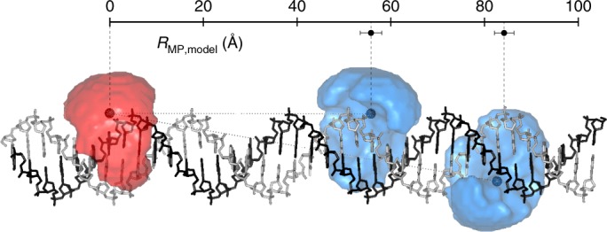
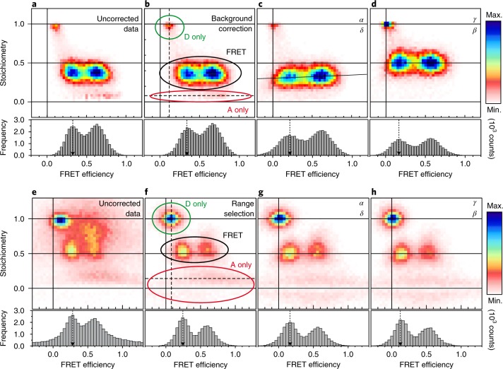
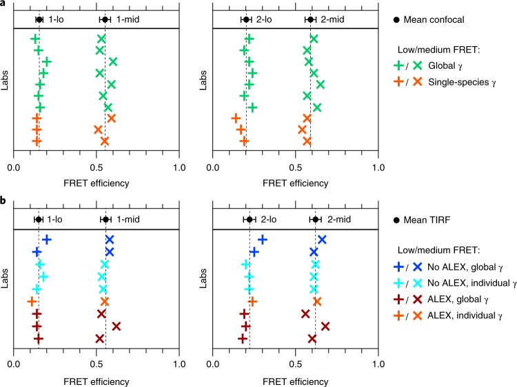
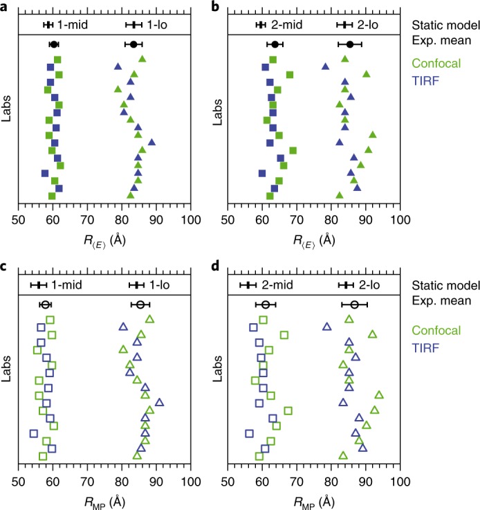
Single-molecule Förster resonance energy transfer (smFRET) is increasingly being used to determine distances, structures, and dynamics of biomolecules in vitro and in vivo. However, generalized protocols and FRET standards to ensure the reproducibility and accuracy of measurements of FRET efficiencies are currently lacking. Here we report the results of a comparative blind study in which 20 labs determined the FRET efficiencies (E) of several dye-labeled DNA duplexes. Using a unified, straightforward method, we obtained FRET efficiencies with s.d. between ±0.02 and ±0.05. We suggest experimental and computational procedures for converting FRET efficiencies into accurate distances, and discuss potential uncertainties in the experiment and the modeling. Our quantitative assessment of the reproducibility of intensity-based smFRET measurements and a unified correction procedure represents an important step toward the validation of distance networks, with the ultimate aim of achieving reliable structural models of biomolecular systems by smFRET-based hybrid methods.


Conflict of interest statement

The authors declare no competing interests.

Figures

Fig. 1
Fig. 1. Schematic of the FRET standard molecules.
Double-stranded DNA was labeled with a FRET pair at 15-bp or 23-bp separation for the “lo” and “mid” samples, respectively (sequences are provided in the Methods). The accessible volumes (AVs) of the dyes (donor, blue; acceptor, red) are illustrated as semi-transparent surfaces and were calculated with freely available software. The mean dye positions are indicated by darker spheres (assuming homogeneously distributed dye positions; Supplementary Note 3). The distance between the mean dye positions is defined as RMP,model. Calculated values for RMP,model and the errors obtained by varying parameters of the AV model are shown (Supplementary Note 3). The B-DNA model was generated with Nucleic Acid Builder version 04/17/2017 for Amber.
Fig. 2
Fig. 2. Stepwise data correction for 1-lo and 1-mid samples.
ad, Workflow for correction of the confocal data for background (ab); leakage (factor α); and direct excitation (δ) (bc), excitation, and detection factors (β, γ) (cd). eh, Workflow for correction of TIRF data for background and photobleaching by selection of the prebleached range (ef); leakage; and direct excitation (fg), detection, and excitation factors (gh). The efficiency histograms show a projection of the data with a stoichiometry between 0.3 and 0.7. The general terms “stoichiometry” and “FRET efficiency” are used in place of the corresponding specific terms for each correction step. Donor (D)-only, FRET, and acceptor (A)-only populations are specified.
Fig. 3
Fig. 3. Summary of the results of the intensity-based methods.
a, Confocal measurements. b, TIRF measurements. Note that some laboratories performed measurements with both methods. The mean ± s.d. is depicted in the upper portion of each plot. Dashed lines indicate mean values (summarized in Supplementary Table 4). Example correction factors are given in Supplementary Table 3.
Fig. 4
Fig. 4. Mean interdye distances determined from 19 〈E〉 values measured in 16 different labs.
a,b, RE for samples 1 (a) and 2 (b). c,d, RMP for samples 1 (c) and 2 (d). Data are shown as individual values (colored symbols) and as the mean (black dots) and s.d., assuming R0 = 62.6 Å and R0 = 68.0 Å for samples 1 and 2, respectively. The black bars at the top of each plot indicate the static model values and their error (determined by variation of model parameters); see Supplementary Table 4 for values. The depicted errors include only the statistical variations of the FRET efficiencies, and do not include the error in the Förster radii; thus these errors represent the precision of the measurement, but not the accuracy. Exp., experimental.
Fig. 5
Fig. 5. Error propagation of experimental uncertainty.
a, RDA uncertainty contributions from the experimental correction factors: ∆Rγ (gamma factor), ΔRbgD and ΔRbgA (background), ∆Rα (leakage), ∆Rδ (direct excitation), and total uncertainty with known R0; crosses indicate the uncertainty of experimental values of RE across the labs. b, Uncertainty in RDA (black line) based on the efficiency-related uncertainty (gray line) and the uncertainty for determining R0 (blue line). Here we used the following uncertainties, which were determined for the confocal-based measurements on sample 1: ΔR0/R0 = 7%, Δγ/γ = 10%, ΔI(BG)/I = 2%, Δα/α = 10%, and Δδ/δ = 10%. Absolute values are presented in Supplementary Table 3.

References

    1. Förster T. Zwischenmolekulare Energiewanderung und Fluoreszenz. Ann. Phys. 1948;437:55–75.
    1. Stryer L, Haugland RP. Energy transfer: a spectroscopic ruler. Proc. Natl Acad. Sci. USA. 1967;58:719–726. - PMC - PubMed
    1. Murchie AI, et al. Fluorescence energy transfer shows that the four-way DNA junction is a right-handed cross of antiparallel molecules. Nature. 1989;341:763–766. - PubMed
    1. Mekler V, et al. Structural organization of bacterial RNA polymerase holoenzyme and the RNA polymerase-promoter open complex. Cell. 2002;108:599–614. - PubMed
    1. Ha T, et al. Probing the interaction between two single molecules: fluorescence resonance energy transfer between a single donor and a single acceptor. Proc. Natl Acad. Sci. USA. 1996;93:6264–6268. - PMC - PubMed

Publication types

MeSH terms