From fix to fit into the autoptic human brains
- PMID: 30173504
- PMCID: PMC6151333
- DOI: 10.4081/ejh.2018.2944
From fix to fit into the autoptic human brains
Abstract
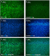
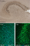
Formalin-fixed, paraffinembedded (FFPE) human brain tissues are very often stored in formalin for long time. Formalin fixation reduces immunostaining, and the DNA/RNA extraction from FFPE brain tissue becomes suboptimal. At present, there are different protocols of fixation and several procedures and kits to extract DNA/RNA from paraffin embedding tissue, but a gold standard protocol remains distant. In this study, we analyzed four types of fixation systems and compared histo and immuno-staining. Based on our results, we propose a modified method of combined fixation in formalin and formic acid for the autoptic adult brain to obtain easy, fast, safe and efficient immunolabelling of long-stored FFPE tissue. In particular, we have achieved an improved preservation of cellular morphology and obtained success in postmortem immunostaining for NeuN. This nuclear antigen is an important marker for mapping neurons, for example, to evaluate the histopathology of temporal lobe epilepsy or to draw the topography of cardiorespiratory brainstem nuclei in sudden infant death syndrome (SIDS). However, NeuN staining is frequently faint or lost in postmortem human brain tissues. In addition, we attained Fluoro Jade C staining, a marker of neurodegeneration, and immunofluorescent staining for stem cell antigens in the postnatal human brain, utilizing custom fit fixation procedures.
Conflict of interest statement
Conflict of interest: The authors received no financial support for the research, authorship, and/or publication of this article.
Figures


Similar articles
-
Will PAXgene substitute formalin? A morphological and molecular comparative study using a new fixative system.J Clin Pathol. 2013 Feb;66(2):124-35. doi: 10.1136/jclinpath-2012-200983. Epub 2012 Nov 3. J Clin Pathol. 2013. PMID: 23125305
-
Immunostaining for NeuN Does Not Show all Mature and Healthy Neurons in the Human and Pig Brain: Focus on the Hippocampus.Appl Immunohistochem Mol Morphol. 2021 Jul 1;29(6):e46-e56. doi: 10.1097/PAI.0000000000000925. Appl Immunohistochem Mol Morphol. 2021. PMID: 33710124
-
An enhanced antigen-retrieval protocol for immunohistochemical staining of formalin-fixed, paraffin-embedded tissues.Methods Mol Biol. 2011;717:101-10. doi: 10.1007/978-1-61779-024-9_6. Methods Mol Biol. 2011. PMID: 21370027
-
Analysis of formalin-fixed, paraffin-embedded (FFPE) tissue via proteomic techniques and misconceptions of antigen retrieval.Biotechniques. 2016 May 1;60(5):229-38. doi: 10.2144/000114414. eCollection 2016. Biotechniques. 2016. PMID: 27177815 Review.
-
A review of preanalytical factors affecting molecular, protein, and morphological analysis of formalin-fixed, paraffin-embedded (FFPE) tissue: how well do you know your FFPE specimen?Arch Pathol Lab Med. 2014 Nov;138(11):1520-30. doi: 10.5858/arpa.2013-0691-RA. Arch Pathol Lab Med. 2014. PMID: 25357115 Review.
Cited by
-
A journal of histochemistry as a forum for non-histochemical scientific societies.Eur J Histochem. 2019 Dec 23;63(4):3106. doi: 10.4081/ejh.2019.3106. Eur J Histochem. 2019. PMID: 31868322 Free PMC article.
-
Effect of the combination of high-frequency repetitive magnetic stimulation and neurotropin on injured sciatic nerve regeneration in rats.Neural Regen Res. 2020 Jan;15(1):145-151. doi: 10.4103/1673-5374.264461. Neural Regen Res. 2020. PMID: 31535663 Free PMC article.
-
In Silico and In Vivo: Evaluating the Therapeutic Potential of Kaempferol, Quercetin, and Catechin to Treat Chronic Epilepsy in a Rat Model.Front Bioeng Biotechnol. 2021 Nov 4;9:754952. doi: 10.3389/fbioe.2021.754952. eCollection 2021. Front Bioeng Biotechnol. 2021. PMID: 34805114 Free PMC article.
-
Are pre-analytical factors fully considered in forensic FFPE molecular analyses? A systematic review reveals the need for standardised procedures.Int J Legal Med. 2025 Jul;139(4):1439-1452. doi: 10.1007/s00414-025-03480-8. Epub 2025 Apr 2. Int J Legal Med. 2025. PMID: 40172636 Free PMC article.
-
Apoptosis in Postmortal Tissues of Goat Spinal Cords and Survival of Resident Neural Progenitors.Int J Mol Sci. 2024 Apr 25;25(9):4683. doi: 10.3390/ijms25094683. Int J Mol Sci. 2024. PMID: 38731901 Free PMC article.
References
-
- Liu JY, Martinian L, Thom M, Sisodiya SM. Immunolabeling recovery in archival, post-mortem, human brain tissue using modified antigen retrieval and the catalyzed signal amplification system. J Neurosci Methods 2010;190:49-56. - PubMed
-
- Carturan E, Tester DJ, Brost BC, Basso C, Thiene G, Ackerman MJ. postmortem genetic testing for conventional autopsy–negative sudden unexplained death: An evaluation of different DNA extraction protocols and the feasibility of mutational analysis from archival paraffin-embedded heart tissue. Am J Clin Pathol 2008;129:391-7. - PMC - PubMed
-
- Marwitz S, Kolarova J, Reck M, Reinmuth N, Kugler C, Schadlich I, et al. The tissue is the issue: improved methylome analysis from paraffinembedded tissues by application of the HOPE technique. Lab Invest 2014;94: 927-33. - PubMed
MeSH terms
Substances
LinkOut - more resources
Full Text Sources
Other Literature Sources

