. 2018 Sep 3;16(1):97.
doi: 10.1186/s12915-018-0561-0.

Microdomain formation is a general property of bacterial membrane proteins and induces heterogeneity of diffusion patterns

Affiliations

Microdomain formation is a general property of bacterial membrane proteins and induces heterogeneity of diffusion patterns

Daniella Lucena et al. BMC Biol. .

Abstract

Background: Proteins within the cytoplasmic membrane display distinct localization patterns and arrangements. While multiple models exist describing the dynamics of membrane proteins, to date, there have been few systematic studies, particularly in bacteria, to evaluate how protein size, number of transmembrane domains, and temperature affect their diffusion, and if conserved localization patterns exist.

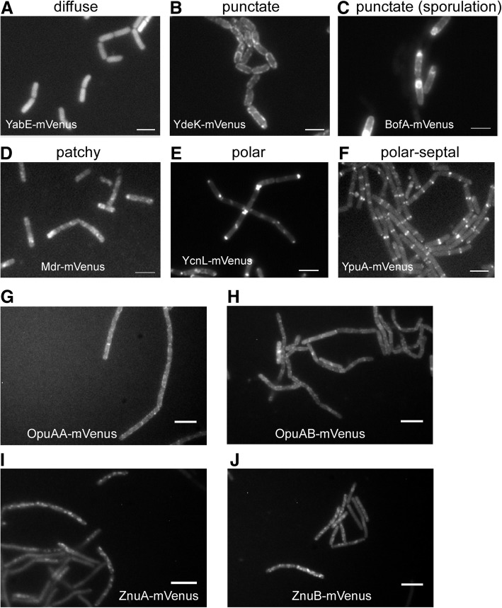
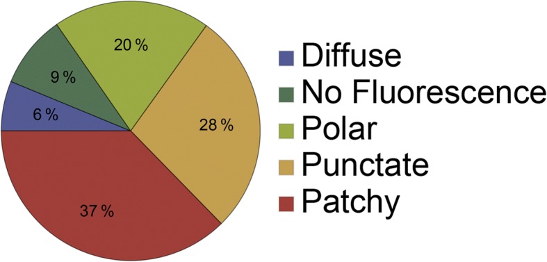
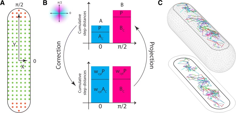
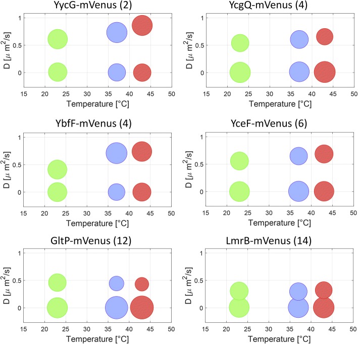
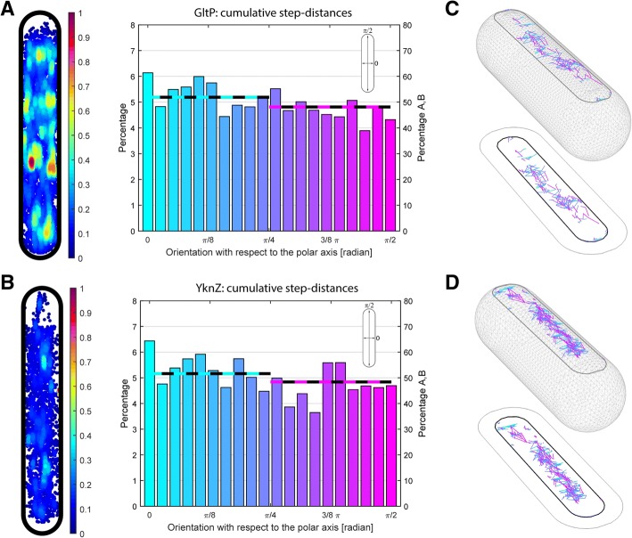
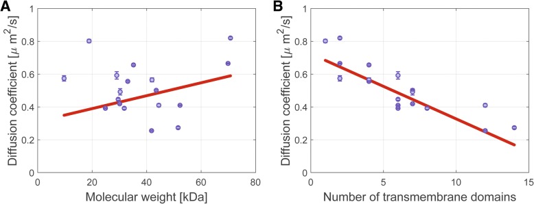
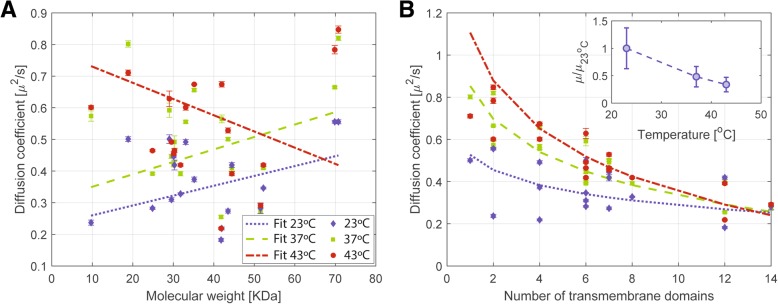
Results: We have used fluorescence microscopy, single-molecule tracking (SMT), and computer-aided visualization methods to obtain a better understanding of the three-dimensional organization of bacterial membrane proteins, using the model bacterium Bacillus subtilis. First, we carried out a systematic study of the localization of over 200 B. subtilis membrane proteins, tagged with monomeric mVenus-YFP at their original gene locus. Their subcellular localization could be discriminated in polar, septal, patchy, and punctate patterns. Almost 20% of membrane proteins specifically localized to the cell poles, and a vast majority of all proteins localized in distinct structures, which we term microdomains. Dynamics were analyzed for selected membrane proteins, using SMT. Diffusion coefficients of the analyzed transmembrane proteins did not correlate with protein molecular weight, but correlated inversely with the number of transmembrane helices, i.e., transmembrane radius. We observed that temperature can strongly influence diffusion on the membrane, in that upon growth temperature upshift, diffusion coefficients of membrane proteins increased and still correlated inversely to the number of transmembrane domains, following the Saffman-Delbrück relation.

Conclusions: The vast majority of membrane proteins localized to distinct multimeric assemblies. Diffusion of membrane proteins can be suitably described by discriminating diffusion coefficients into two protein populations, one mobile and one immobile, the latter likely constituting microdomains. Our results show there is high heterogeneity and yet structural order in the cell membrane, and provide a roadmap for our understanding of membrane organization in prokaryotes.

Keywords: B. subtilis; Diffusion; Membrane dynamics; Membrane protein localization; Single-molecule tracking.


Conflict of interest statement

Ethics approval and consent to participate

Not applicable.

Consent for publication

Not applicable.

Competing interests

The authors declare that they have no competing interests.

Publisher’s Note

Springer Nature remains neutral with regard to jurisdictional claims in published maps and institutional affiliations.

Figures

Fig. 1
Fig. 1
Representative pictures of fluorescence microscopy experiments showing patterns of membrane protein distribution according to their subcellular localization. The mVenus-tagged membrane proteins were observed in a diffuse, b punctate, c punctate/sporulation, d patchy, e polar, and f polar/septal patterns of distribution. g, h OpuAA and OpuAB form a complex in vivo, and both localize in a punctate pattern. i, j Both subunits of the ZnuAB complex localize in a patchy pattern. Gray lines in panels af 2 μm, white lines in gj 3 μm
Fig. 2
Fig. 2
Pie chart representing membrane protein distribution according to their subcellular localization. For 15% of the proteins tested, there was no membrane localization, either diffuse (6%) or no fluorescence (9%) was observed. The remaining proteins were localized laterally on the membrane in a punctate fashion (28%), in a patchy distribution (37%), or at the cell poles and/or septa (20%)
Fig. 3
Fig. 3
Curvature correction factor. In a, a schematic representation of a B. subtilis average cell aligned to coordinate axes. The polar and cylindrical regions are represented by orange and green, respectively. b Projection of curvature on observation plane impairs equality between components of cumulative step-distances along x (cyan) and y (magenta) axes. P (P) and Ac (Bc) are the cumulative step-distances observed in the polar and cylindrical regions along short (long) axis. Equality can be restored by properly weighing these values by the factors wcyl = Bc/Ac and w = B/A, from which the curvature correction coefficient can be obtained (see Methods). c A realistic three-dimensional representation of tracks of the protein YbfF show that in our set-up, the curvature effect is not too pronounced. The region accessible with our depth of focus is delimited by the black curve. On the xy plane, we show the projections of observed boundary (gray), whole cell boundary (black), and the observed tracks
Fig. 4
Fig. 4
Variation of diffusion coefficients on dependence of growth temperature and number of transmembrane domains (shown in brackets). Graphs show two diffusing populations of mVenus-tagged proteins in B. subtilis PY 79 cultures grown at 23 °C, 37 °C, or 43 °C. The size of the circles corresponds to the percentage of molecules in each fraction. The number of transmembrane domains of each protein is given between parentheses
Fig. 5
Fig. 5
Density map and directionality histogram for proteins a GltP and b YknZ. The left panel shows density maps of the tracks in the average size of the bacterial cell from each protein database. In the histograms, the orientation of the tracks was calculated with respect to the short axis of the bacterial cells. The left y axis refers to the values of the cyan and magenta lines, which represent the cumulative step-distances along the short and long axis, respectively. Cumulative step-distances are the sum of all magnitudes of the components along the coordinate axes of all distances of every track at every time. The right y axis of the histograms shows the percentage for a finer division in smaller angles (20 bins). Distances were corrected for curvature as explained in the “Methods” section. c, d 3D visualization of tracks for two representative cells from the GltP and YknZ databases, respectively. The region accessible with our depth of focus is delimited by the black curve. Tracks are sorted according to their directions with the same color codes as in a and b
Fig. 6
Fig. 6
Dependence of membrane protein diffusion coefficients on protein size or on the number of transmembrane domains. Diffusion coefficients of mVenus fusion membrane proteins are plotted either a against protein molecular weight or b against the number of transmembrane domains. Linear trend lines are shown in red
Fig. 7
Fig. 7
Dependence of membrane protein diffusion coefficients on temperature. Diffusion coefficients of selected mVenus fusion proteins were plotted against their molecular weight (a) and the number of transmembrane domains (TMs) (b). The data is fitted in a to a linear relation (for comparison) and in b to the Saffmann–Delbrück model. Dotted blue lines, dashed green lines, and dash-dotted red lines refer to 23 °C, 37 °C, and 43 °C, respectively. Inset in b shows the relative decrease of membrane viscosity as a function of temperature

References

    1. Shapiro L, McAdams HH, Losick R. Why and how bacteria localize proteins. Science. 2009;326(5957):1225–1228. doi: 10.1126/science.1175685. - DOI - PMC - PubMed
    1. Manzo C, Garcia-Parajo MF. A review of progress in single particle tracking: from methods to biophysical insights. Rep Prog Phys. 2015;78(12):124601. doi: 10.1088/0034-4885/78/12/124601. - DOI - PubMed
    1. Oswald F, Varadarajan A, Lill H, Peterman EJ, Bollen YJ. MreB-dependent organization of the E. coli cytoplasmic membrane controls membrane protein diffusion. Biophys J. 2016;110(5):1139–1149. doi: 10.1016/j.bpj.2016.01.010. - DOI - PMC - PubMed
    1. Jackson D, Wang X, Rudner DZ. Spatio-temporal organization of replication in bacteria and eukaryotes (nucleoids and nuclei) Cold Spring Harb Perspect Biol. 2012;4(8):a010389. - PMC - PubMed
    1. Hofling F, Franosch T. Anomalous transport in the crowded world of biological cells. Rep Prog Phys. 2013;76(4):046602. doi: 10.1088/0034-4885/76/4/046602. - DOI - PubMed

Publication types

MeSH terms

LinkOut - more resources