Review
. 2018 Nov:71:43-55.
doi: 10.1016/j.dnarep.2018.08.006. Epub 2018 Aug 23.

Structural basis of DNA lesion recognition for eukaryotic transcription-coupled nucleotide excision repair

Affiliations
Review

Structural basis of DNA lesion recognition for eukaryotic transcription-coupled nucleotide excision repair

Wei Wang et al. DNA Repair (Amst). 2018 Nov.

Abstract

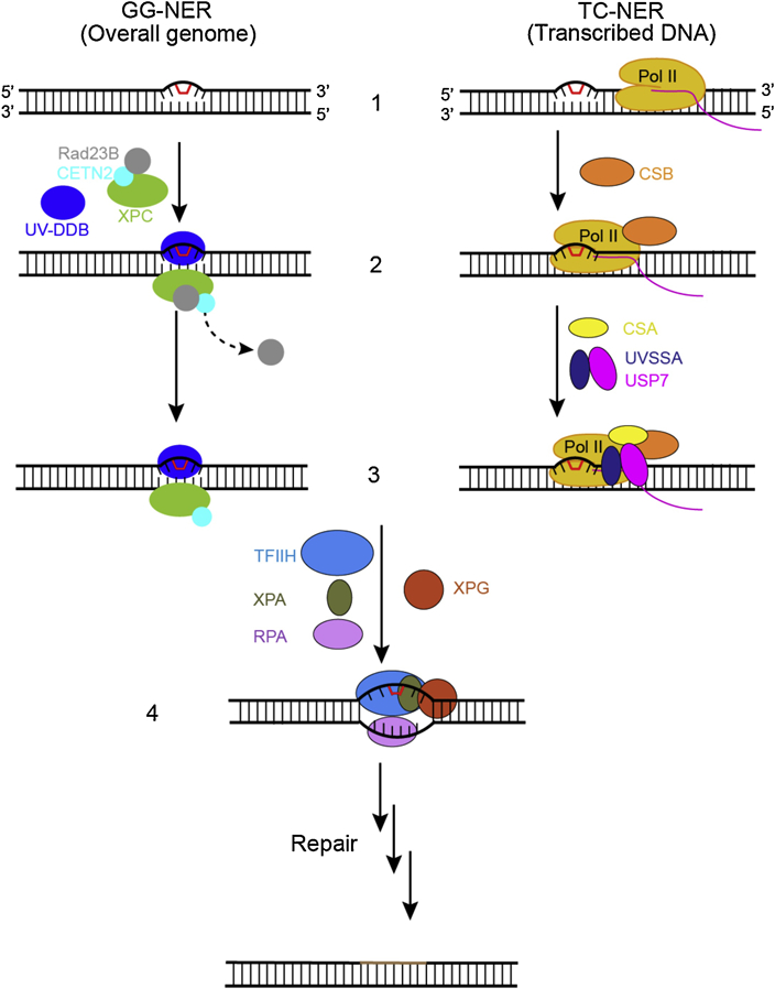
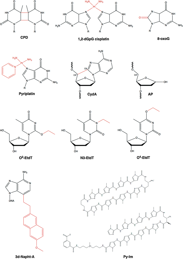
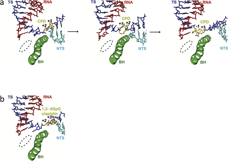
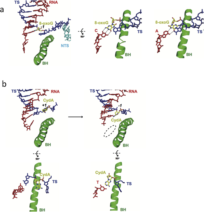
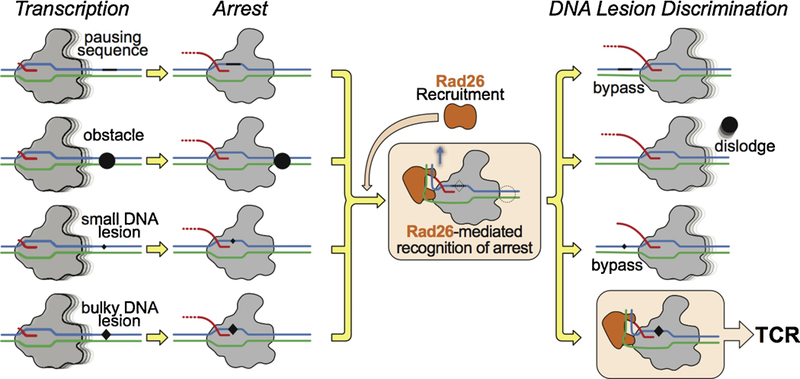
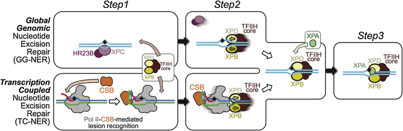
Eukaryotic transcription-coupled nucleotide excision repair (TC-NER) is a pathway that removes DNA lesions capable of blocking RNA polymerase II (Pol II) transcription from the template strand. This process is initiated by lesion-arrested Pol II and the recruitment of Cockayne Syndrome B protein (CSB). In this review, we will focus on the lesion recognition steps of eukaryotic TC-NER and summarize the recent research progress toward understanding the structural basis of Pol II-mediated lesion recognition and Pol II-CSB interactions. We will discuss the roles of CSB in both TC-NER initiation and transcription elongation. Finally, we propose an updated model of tripartite lesion recognition and verification for TC-NER in which CSB ensures Pol II-mediated recognition of DNA lesions for TC-NER.

Keywords: Cockayne syndrome; DNA damage; Lesion recognition; Nucleotide excision repair; RNA polymerase II; Transcription-coupled nucleotide excision repair; Transcriptional arrest.


Conflict of interest statement

Conflict of Interest

The authors declare no conflict of interest.

Figures

Figure 1.
Figure 1.
The recognition steps of two subpathways of nucleotide excision repair. In the global genome nucleotide excision repair subpathway (GG-NER), left, the damage sensor XPC, in complex with UV excision repair protein RAD23 homologue B (RAD23B) and centrin 2 (CETN2), which binds the non-damaged strand opposite the lesion with the help of the UV– DDB complex (step 2, left). Binding of the XPC complex to the damaged site results in RAD23B dissociation from the complex (step 3, left). In the transcription-coupled subpathway (TC-NER), damage recognition is initiated by the stalling of RNA polymerase II (Pol II). The stalled Pol II recruits CSB (step 2, right) and the Pol II–CSB complex serves as a platform to further recruitment of downstream repair factors such as CSA and UVSSA-USP7 (step 3, right). After damage recognition (step1–3), the TFIIH complex is recruited to the lesion in both GG-NER and TC-NER, along with XPA, RPA, and XPG (step 4). Once DNA lesion is verified, the damage-containing DNA short fragment is removed via dual incisions, new DNA fragment is synthesized, and NER reaction is completed through sealing the final nick by DNA ligase.
Figure 2.
Figure 2.
Chemical structures of DNA lesions: cyclobutane pyrimidine dimer (CPD), 1,2- dGpG cisplatin, 8-oxoguanine (8-oxoG), monofunctional pyriplatin, 8,5′-cyclo-2′-deoxyadenosine (CydA), abasic site (AP), O2-, N3- and O4-ethylthymideine (O2-, N3, and O4-EtdT), 3-deaza-3-methoxynaphtylethyl-adenosine (3d-Napht-dA), and non-covalent DNA binders Pyrrole-imidazole (Py-Im) polyamides. The DNA lesion is highlighted in red.
Figure 3.
Figure 3.
The structures of Pol II EC stalled at intrastrand crosslinks. a, CPD lesion-induced Pol II stall complex (PDB IDs: 2JA6, 4A93 and 2JA7). b, 1,2-dGpG cisplatin DNA lesion-induced Pol II stall complex (PDB ID: 2R7Z). RNA, template strand, non-template strand, and the bridge helix motif of RNA Pol II (Rpb1 810–840) are shown in red, blue, cyan, and green, respectively. The DNA lesion are highlighted and labeled in yellow. The template registers are also marked in the figure. The Pol II active site is indicated in dashed circle.
Figure 4.
Figure 4.
RNA Pol II recognition of oxidative DNA lesions. a. Pol II recognition of 8-oxoguanine (8-oxoG) lesion. Canonical Watson-Crick pair is formed between 8-oxo dG and C (PDB ID: 3I4M), but 8-oxo dG adopts syn-form when pairing with adenine in the Pol II-active site (PDB ID: 3I4N). b. Pol II EC stalled at an 8,5′-cyclo-2′-deoxyadenosine (CydA) site (PDB IDs: 4X6A and 4X67). Color codes are the same as Figure 3.
Figure 5.
Figure 5.
Pol II EC stalled by major groove or minor groove obstructions. a. Pol II EC stalled by a pyriplatin-DNA monofunctional adduct (PDB IDs: 3M4O and 3M3Y). b. Structural modeling of RNA Pol II transcriptional pausing by Py-Im polyamide. Two critical residues (R1386 and H1387) from the conserved Switch 1 region of Rpb1 play a key role in slowing down transcription progress through the polyamide-bound minor groove step by step. c. The Pol II EC adopts the post-translocation state with the modified adenine base (3d-Napht-dA) at +1 position, with 3-deaza-3-methoxynaphtylethyl group contacting the bridge helix in the Pol II active center from underneath (PDB ID: 5OT2). Color codes are the same as Figure 3.
Figure 6.
Figure 6.
Structural modeling showed distinct effects of O4- and O2-EtdT lesions on transcriptional bypass. Briefly, the alkylation in the major groove (O4-EtdT) hardly affects translocation, whereas the minor-groove alkylation (O2-EtdT) has strong steric clash with the P448 residue on the ‘Pro-gate loop’ during translocation. Color codes are the same as Figure 3.
Figure 7.
Figure 7.
Structural analysis of transcription stalling by the AP site at insertion and extension steps with four structural snapshots (PDB IDs: 6BLO, 6BLP, 6BM2 and 6BM4). Color codes are the same as Figure 3.
Figure 8.
Figure 8.
Rad26 helps Pol II discriminate among different transcription obstacles a-c.Transcription assays probing the ability of Rad26, Rad26 mutant, and TFIIS to discriminate among three representative transcription obstacles encountered by Pol II: A pause-inducing repetitive A-tract sequence (a); A sequence-specific DNA-binding polyamide (Py-Im) (b); and a TT cyclobutane pyrimidine dimer (CPD) DNA lesion (c). The asterisk in (a) represents a Pol II pausing site upstream of the A-tract sequence. Last lane in (c): full-length transcript in the absence of the CPD lesion.
Figure 9.
Figure 9.
Pol II-CSB structure and upstream DNA bending. a, Atomic model for the Pol II– Rad26 complex. Cartoons of the structures highlighting their orientations. b, Superposition of the scaffolds from the Pol II–Rad26 and Pol II EC structures, with the latter shown in darker colors.
Figure 10.
Figure 10.
Comparison of three stages of Pol II complexes. The initiation open complex structure is shown on the left, PDB ID: 5FYW; The transcription elongation complex is shown in the middle, PDB ID: 5OIK; The transcription repair initiation complex is shown on the right, PDB ID: 5VVR. In the three different stages, TFIIE, Spt4/5, and CSB bind to similar positions on Pol II. Pol II is in gray and other factors are highlighted and labeled as indicated. Template and non-template DNA, and RNA are shown in blue, green, and red, respectively.
Figure 11.
Figure 11.
Model for lesion recognition by CSB. Rad26 recognizes a stalled Pol II and can reduce its dwell time by preventing backtracking, promoting Pol II forward translocation on non-damaged templates, and increasing the chances of transcriptional bypass through less bulky DNA lesions, all of which stimulate transcription elongation. However, Rad26 fails to promote efficient transcriptional bypass of bulky DNA lesions that lead to strong blockage of translocation (such as CPD lesions). The interaction between Rad26 and a Pol II persistently arrested at a bulky lesion would lead to the initiation of TCR.
Figure 12.
Figure 12.
Three DNA lesion checkpoints for GG-NER and TC-NER. Check step 1: For GG-NER, XPC/HR23B detects base-pair disruption and helix distortion and binds to the DNA strand opposite to that carrying the lesion. This constitutes the initial lesion recognition. For TCR, CSB is recruited to a stalled Pol II to discriminate genuine DNA lesion-induced transcription arrest from other forms of transcriptional arrest, as diagrammed in panel a. At this step, CSB acts in conjunction with Pol II to mediate the initial recognition of DNA lesions that block transcription translocation. Check step 2: Core TFIIH is recruited to further verify the DNA lesion. In GG-NER, the XPD and XPB helicases in core TFIIH translocate the complex towards the lesion. This is the result of XPD tracking along the damage-containing strand in a 5´ to 3´ direction and XPB tracking along the opposite strand (non-damaged) in a 3´ to 5´ direction. In TCR, TFIIH is loaded downstream of the arrested Pol II-CSB complex, with XPD and XPB tracking the template and non-template strands, respectively. The XPD/XPB helicases in core TFIIH translocate towards the lesion, as is the case for GG-NER. As a result, Pol II-CSB is pushed upstream by TFIIH to expose the DNA lesion. Check step 3: XPA is recruited for a final validation of the TFIIH-recognized lesion and to ensure that only genuine NER lesions are subjected to dual incision by endonucleases ERCC1/XPF and XPG and downstream repair synthesis.

References

    1. Sancar A & Reardon JT (2004) Nucleotide excision repair in E. coli and man. Adv Protein Chem 69:43–71. - PubMed
    1. Hanawalt PC & Spivak G (2008) Transcription-coupled DNA repair: two decades of progress and surprises. Nat Rev Mol Cell Biol 9(12):958–970. - PubMed
    1. Spivak G (2015) Nucleotide excision repair in humans. DNA Repair (Amst) 36:13–18. - PMC - PubMed
    1. Bohr VA, Smith CA, Okumoto DS, & Hanawalt PC (1985) DNA repair in an active gene: removal of pyrimidine dimers from the DHFR gene of CHO cells is much more efficient than in the genome overall. Cell 40(2):359–369. - PubMed
    1. Mellon I, Spivak G, & Hanawalt PC (1987) Selective removal of transcription-blocking DNA damage from the transcribed strand of the mammalian DHFR gene. Cell 51(2):241–249. - PubMed

Publication types

LinkOut - more resources