. 2018 Sep 11;11(3):727-740.
doi: 10.1016/j.stemcr.2018.08.003. Epub 2018 Aug 30.

Organoids from Nephrotic Disease-Derived iPSCs Identify Impaired NEPHRIN Localization and Slit Diaphragm Formation in Kidney Podocytes

Affiliations

Organoids from Nephrotic Disease-Derived iPSCs Identify Impaired NEPHRIN Localization and Slit Diaphragm Formation in Kidney Podocytes

Shunsuke Tanigawa et al. Stem Cell Reports. .

Abstract

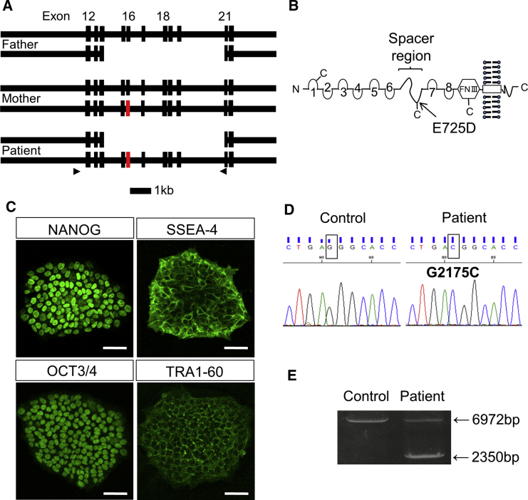
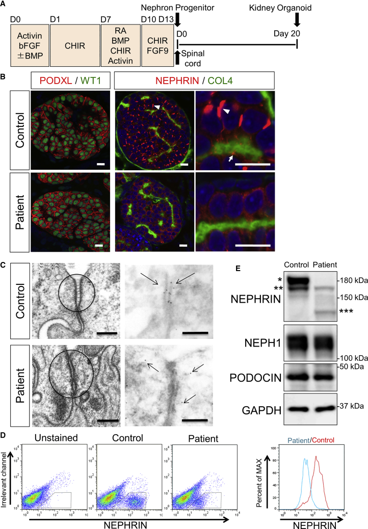
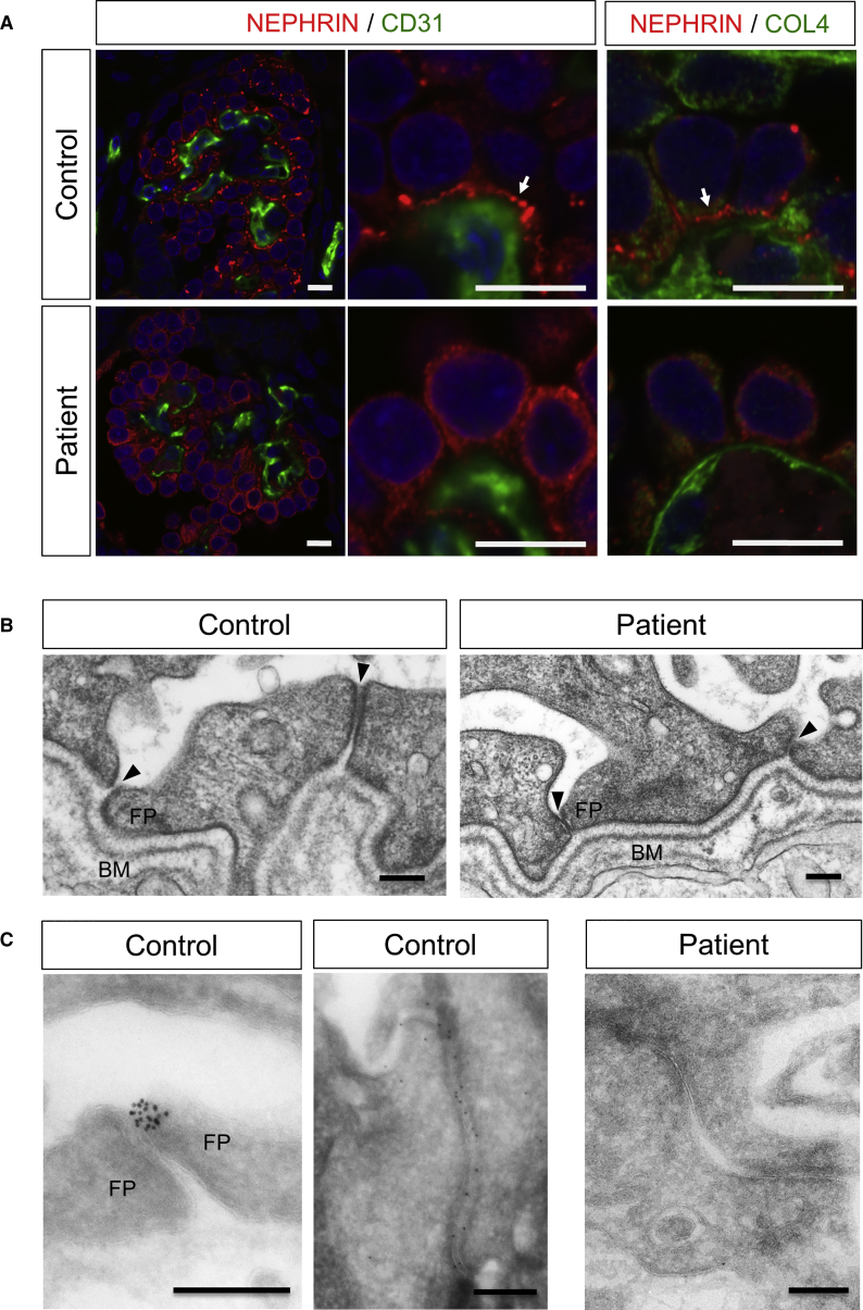
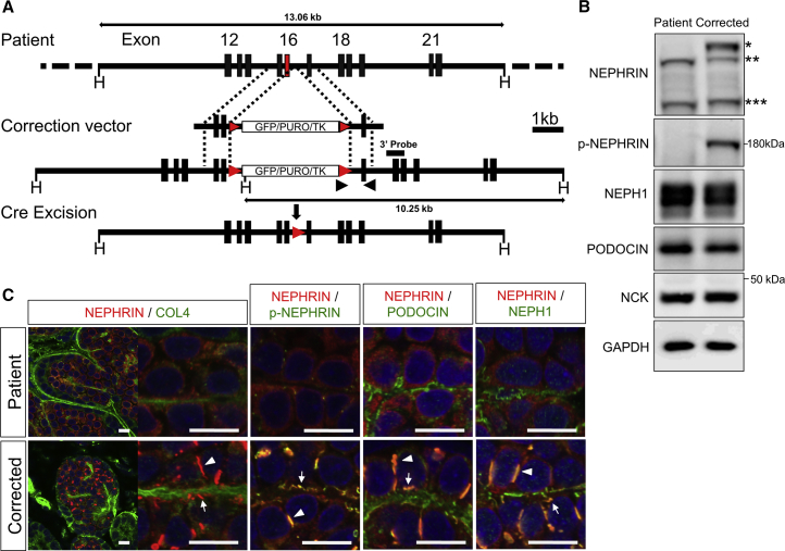
Mutations in the NPHS1 gene, which encodes NEPHRIN, cause congenital nephrotic syndrome, resulting from impaired slit diaphragm (SD) formation in glomerular podocytes. However, methods for SD reconstitution have been unavailable, thereby limiting studies in the field. In the present study, we established human induced pluripotent stem cells (iPSCs) from a patient with an NPHS1 missense mutation, and reproduced the SD formation process using iPSC-derived kidney organoids. The mutant NEPHRIN failed to become localized on the cell surface for pre-SD domain formation in the induced podocytes. Upon transplantation, the mutant podocytes developed foot processes, but exhibited impaired SD formation. Genetic correction of the single amino acid mutation restored NEPHRIN localization and phosphorylation, colocalization of other SD-associated proteins, and SD formation. Thus, these kidney organoids from patient-derived iPSCs identified SD abnormalities in the podocytes at the initial phase of congenital nephrotic disease.

Keywords: NEPHRIN; NPHS1; iPSCs; kidney; nephrotic syndrome; podocyte; slit diaphragm.


Figures

None
Graphical abstract
Figure 1
Figure 1
Establishment of iPSCs from a Patient with NPHS1 Mutations (A) Exon/intron structures of the paternal, maternal, and patient alleles of NPHS1. A point mutation (G2175C) is located at exon 16 of the maternal allele (red). The arrowheads show the primers used for the experiment shown in (E). Scale bar, 1 kb. (B) Putative NEPHRIN protein produced from the maternal allele. The arrow shows the amino acid substitution location in the spacer region. FNIII, fibronectin-like domain III. (C) Stem cell markers expressed in the patient-derived iPSCs. Scale bars, 50 μm. (D) NPHS1 sequences in the control and patient-derived iPSCs. The rectangular boxes show the nucleotide mutation at position 2175. (E) Deletion of the paternal allele, as confirmed by genomic PCR. The primers shown in (A) were used. See also Figure S1.
Figure 2
Figure 2
Point Mutation Impairs Protein Processing toward the Cell Surface (A) Flow cytometry analysis of HEK293 cells overexpressing wild-type (WT) or mutant (MT) NEPHRIN using an antibody (48E11) against the extracellular domain of NEPHRIN. The y axis shows the cell counts normalized to 100% of the total cells. Blue, unstained HEK293 cells; red, tetracycline-induced HEK293 cells. (B) Immunostaining of HEK293 cells overexpressing WT or MT NEPHRIN using an antibody (50A9) against the extracellular domain of NEPHRIN in the absence of detergent. Note the reduced signals for MT NEPHRIN. Wheat germ agglutinin (WGA) binds to the surface glycoproteins. Scale bars, 20 μm. (C) Biotin-mediated cell surface NEPHRIN labeling in HEK293 cells. TfR, transferrin receptor (negative control). Biotinylated cell surface components were isolated with streptavidin-Sepharose and isolated proteins were detected by immunoblotting with the anti-NEPHRIN antibody that recognizes the cytoplasmic region of NEPHRIN (Bio/Strep-IP). Input represents 0.2% of the total protein used for analysis. (D) Endoglycosidase H (endo H)-mediated cleavage of the lower NEPHRIN bands. Lysates of HEK293 cells overexpressing human WT or MT NEPHRIN were incubated with or without endo H. Endo H induced loss of the lower NEPHRIN band and appearance of a faster migrating band. The upper band of WT NEPHRIN was unaffected. (E) Kinetics of NEPHRIN on the cell surface of HEK293 cells after tetracycline treatment. Cells were harvested at 0, 6, 24, and 48 hr, and cell surface components were detected by biotin-mediated labeling as described in (C). See also Figure S2.
Figure 3
Figure 3
Mutant Podocytes Exhibit Reduced Cell Surface Localization of NEPHRIN (A) Outline of the protocol to induce podocytes from iPSCs through nephron progenitors. CHIR, CHIR99021; RA, retinoic acid. Spinal cord was attached to nephron progenitors at day 13 to induce nephrogenesis, and the kidney organoids were cultured for a further 20 days. (B) Section immunostaining of kidney organoids at day 20 after nephron induction, showing altered NEPHRIN localization in the patient-derived podocytes. The arrowheads point to the lateral pre-SD domains and the arrows point to the basal pre-SD domains. PODXL, PODOCALYXIN; COL4, type IV collagen. Scale bars, 10 μm. (C) Electron microscopic images of podocytes in the kidney organoids. Left panels: ladder-like structures are formed in the control podocytes only. Right panels: immunoelectron microscopic images showing NEPHRIN localization at the intercellular junctions only in the control podocytes. The arrows show the NEPHRIN signals. Scale bars, 200 nm. (D) Flow cytometry analysis of NEPHRIN in kidney organoids using an antibody (48E11) against the extracellular domain of NEPHRIN. The histograms show cell counts normalized to 100% of the total cells in the NEPHRIN+ gates. (E) Western blots of NEPHRIN and SD-associated proteins from kidney organoids. The upper band detected only in the control; ∗∗lower bands detected both in the control and the mutant; and ∗∗∗the additional band presumably representing the paternal truncated protein. See also Figures S1 and S5.
Figure 4
Figure 4
Mutant Podocytes Form Foot Processes, but Exhibit Impaired SD Formation (A) Immunostaining of iPSC-derived glomeruli and podocytes after transplantation. Glomeruli are vascularized with CD31+ endothelial cells in both groups, but the NEPHRIN distributions are different. The arrows point to the basal linear expression of NEPHRIN. COL4, type IV collagen. Scale bars, 10 μm. (B) Widened filtration slits in the control podocytes, but not in mutant podocytes, as shown by electron microscopy. The right junction in the control is still at the pre-SD stage. The arrowheads show filtration slits. BM, basement membrane; FP, foot process. Scale bars, 200 nm. (C) Failure of NEPHRIN localization between the foot processes (FP), as shown by immunoelectron microscopy. The control panels show two independent regions of filtration slits (left, slit between foot processes; right, pre-SD stage). Scale bars, 200 nm. See also Figure S3.
Figure 5
Figure 5
Genetic Correction of the Point Mutation Restores NEPHRIN Localization (A) Correction of the point mutation by homologous recombination, followed by Cre-mediated removal of the selection cassette. The puromycin-resistance cassette (GFP/PURO/TK) is flanked by loxP sites (red arrowheads). Black arrowheads show the primers used for screening. Scale bar, 1 kb. (B) Western blots of NEPHRIN and SD-associated proteins from the kidney organoids in vitro (day 20). The upper band detected only in the corrected podocytes; ∗∗lower bands detected in both samples; and ∗∗∗the additional bands presumably representing the paternal truncated protein. (C) Restored localization and phosphorylation of NEPHRIN in the kidney organoids in vitro (day 20). NEPHRIN colocalizes with p-NEPHRIN, PODOCIN, and NEPH1 in the genetically corrected organoids. The arrowheads point to the lateral pre-SD domains and the arrows point to the basal pre-SD domains. Scale bars, 10 μm. See also Figures S1 and S4.
Figure 6
Figure 6
Genetic Correction Restores the SD Formation Process (A) Immunoelectron microscopic images showing the restored NEPHRIN localization (arrows) in the intercellular regions of the podocytes in the corrected kidney organoids in vitro (day 20). Scale bars, 200 nm. (B) Transmission electron microscopic images showing filtration slits in the podocytes after transplantation (day 20). While the patient foot processes are tightly adhered, the corrected foot processes exhibit widened gaps. The arrowheads show filtration slits. BM, basement membrane; FP, foot process; EC, endothelial cells. Scale bars, 200 nm.

References

    1. Bantounas I., Ranjzad P., Tengku F., Silajdžić E., Forster D., Asselin M.C., Lewis P., Lennon R., Plagge A., Wang Q. Generation of functioning nephrons by implanting human pluripotent stem cell-derived kidney progenitors. Stem Cell Reports. 2018;10:766–779. - PMC - PubMed
    1. Beltcheva O., Martin P., Lenkkeri U., Tryggvason K. Mutation spectrum in the nephrin gene (NPHS1) in congenital nephrotic syndrome. Hum. Mutat. 2001;17:368–373. - PubMed
    1. Chittiprol S., Chen P., Petrovic-Djergovic D., Eichler T., Ransom R.F. Marker expression, behaviors, and responses vary in different lines of conditionally immortalized cultured podocytes. Am. J. Physiol. Renal Physiol. 2011;301:F660–F671. - PMC - PubMed
    1. Colls P., Blanco J., Martínez-Pasarell O., Vidal F., Egozcue J., Márquez C., Guitart M., Templado C. Chromosome segregation in a man heterozygous for a pericentric inversion, inv(9)(p11q13), analyzed by using sperm karyotyping and two-color fluorescence in situ hybridization on sperm nuclei. Hum. Genet. 1997;99:761–765. - PubMed
    1. Donoviel D.B., Freed D.D., Vogel H., Potter D.G., Hawkins E., Barrish J.P., Mathur B.N., Turner C.A., Geske R., Montgomery C.A. Proteinuria and perinatal lethality in mice lacking NEPH1, a novel protein with homology to NEPHRIN. Mol. Cell. Biol. 2001;21:4829–4836. - PMC - PubMed

Publication types