Case Reports
. 2018 Aug 2:2018:3870640.
doi: 10.1155/2018/3870640. eCollection 2018.

Why Asplenic Patients Should Not Take Care of the Neighbour's Dog? A Fatal Course of Capnocytophaga canimorsus Sepsis

Affiliations
Case Reports

Why Asplenic Patients Should Not Take Care of the Neighbour's Dog? A Fatal Course of Capnocytophaga canimorsus Sepsis

Patrick Langguth et al. Case Rep Infect Dis. .

Abstract

Capnocytophaga canimorsus (CC) belongs to the family Flavobacteriaceae which physiologically occurs in the natural flora of the oral mucosa of dogs and cats. In patients with a compromised immune system, CC can induce a systemic infection with a fulminant course of disease. Infections with CC are rare, and the diagnosis is often complicated and prolonged. We describe a patient with a medical history of prior splenectomy who presented with an acute sepsis and disseminated intravascular coagulation (DIC) and was initially treated on Waterhouse-Friderichsen syndrome (WFS). After the patient had died despite forced treatment in the intermediate care unit, the differential diagnosis of CC was confirmed by culture of blood smears. Later on, a retrospective third-party anamnesis revealed that the patient had contact to his neighbour's dog a few days before disease onset. In conclusion, patients with CC infection can mimic WFS and therefore must be included in the differential diagnosis, especially in patients with a corresponding medical history of dog or cat bites, scratches, licks, or simple exposure.


Figures

Figure 1
Figure 1
Contrast-enhanced abdominal CT demonstrates haemorrhage of the swollen adrenal glands (arrow) and status after splenectomy (a). There is fluid collection in the perirenal space (arrowheads), and the corticomedullary differentiation of the kidneys is lost (b).
Figure 2
Figure 2
Cranial CT reveals hyperdense sedimentations (Hounsfield unit (HU) 23) in both posterior lateral ventricles, suggesting haemorrhage or pus.
Figure 3
Figure 3
Necrosis of the left hand.
Figure 4
Figure 4
Haemorrhagic necrosis of the adrenal gland and complete necrosis of the adrenal gland. The different zones of the adrenal cortex cannot be clearly distinguished. Especially the cortex shows extensive haemorrhage (arrow). The cells are ill-defined and intermingled with fibrin exudation, showing nuclear shadows (insert, double arrow). The cellular compartment of the adrenal medulla is necrotic with intermingled fibrin exudation (star) (haematoxylin and eosin, original magnification 4x insert, original magnification 20x).
Figure 5
Figure 5
Dilated vessels, disseminated haemorrhage, and oedema of the brain. The brain shows ubiquitously oedema with loosened parenchyma. The vessels are dilated (arrows). Diffuse erythrocyte extravasation is shown (double arrows; haematoxylin and eosin, original magnification 4x insert, original magnification 20x).
Figure 6
Figure 6
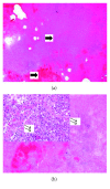
Complete necrosis of the lung with extensive haemorrhage and absceding bronchopneumonia. (a) Completely necrotic lung parenchyma with extensive intra-alveolar haemorrhage. The lung parenchyma is visible as a shadow. (b) An intra-alveolar infiltration by neutrophilic granulocytes (double arrows), focally with abscess formation (haematoxylin and eosin, original magnification 4x insert, original magnification 20x).

References

    1. Bobo R. A., Newton E. J. A previously undescribed gram-negative bacillus causing septicemia and meningitis. American Journal of Clinical Pathology. 1976;65(4):564–569. doi: 10.1093/ajcp/65.4.564. - DOI - PubMed
    1. Janda J. M., Graves M. H., Lindquist D., Probert W. S. Diagnosing Capnocytophaga canimorsus infections. Emerging Infectious Diseases. 2006;12(2):340–342. doi: 10.3201/eid1202.050783. - DOI - PMC - PubMed
    1. Manfredi P., Pagni M., Cornelis G. R. Complete genome sequence of the dog commensal and human pathogen Capnocytophaga canimorsus strain 5. Journal of Bacteriology. 2011;193(19):5558–5559. doi: 10.1128/jb.05853-11. - DOI - PMC - PubMed
    1. Brenner D. J., Hollis D. G., Fanning G. R., Weaver R. E. Capnocytophaga canimorsus sp. nov. (formerly CDC group DF-2), a cause of septicemia following dog bite, and C. cynodegmi sp. nov., a cause of localized wound infection following dog bite. Journal of Clinical Microbiology. 1989;27(2):231–235. - PMC - PubMed
    1. Lion C., Escande F., Burdin J. C. Capnocytophaga canimorsus infection in human: review of the literature and cases report. European Journal of Epidemiology. 1996;12(5):521–533. doi: 10.1007/bf00144007. - DOI - PubMed

Publication types

LinkOut - more resources