

Simultaneous bilateral pneumothorax in an immunocompromised HIV patient with Pneumocystis jirovecii pneumonia
- PMID: 30175036
- PMCID: PMC6115535
- DOI: 10.1016/j.rmcr.2018.08.008
Simultaneous bilateral pneumothorax in an immunocompromised HIV patient with Pneumocystis jirovecii pneumonia
Abstract
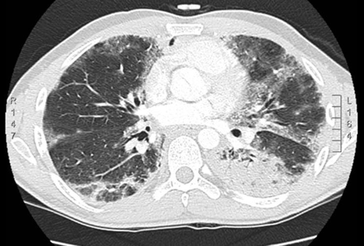
Pneumocystis in humans is caused by a unicellular and eukaryotic organism called P. jirovecii. The overall incidence of P. jirovecii pneumonia (PCP) has decreased with the use of highly active antiretroviral therapy and the use of chemoprophylaxis with trimethroprim sulfametoxazole (TMP/SMX) in cases of immunosuppressed patients. However, approximately 85% of patients with advanced HIV infections continue to experience this disease with inadequate management. Pneumocystis infection can present with spontaneous pneumothorax in 2-6% of cases [8] which can be a potentially fatal complication. We report the case of a 32-year-old man presented with P. jirovecii pneumonia who developed cystic lesions and spontaneous bilateral pneumothorax in spite of TMP/SMX treatment. We consider it an interesting clinical case because few simultaneous bilateral pneumothorax cases have been described directly related to the PCP.
Figures


References
-
- Salzer H.J.F., Schäfer G., Hoenigl M., Günther G., Hoffmann C., Kalsdorf B., Alanio A., Lange C. Clinical, diagnostic, and treatment disparities between HIV-infected and non-HIV-infected immunocompromised patients with pneumocystis jiroveci pneumonia. Respiration. 2018;96(1):52–65. - PubMed
-
- Park Y.K., Jung H.C., Kim S.Y., Kim M.Y., Jo K., Kim S.Y., Kang B., Woo G., Choi H.J., Wie S.H. Spontaneous pneumomediastinum, pneumopericardium, and pneumothorax with respiratory failure in a patient with AIDS and pneumocystis jirovecii pneumonia. Infect. Chemother. 2014 Sep;46(3):204–208. - PMC - PubMed
Publication types
LinkOut - more resources
Full Text Sources
Other Literature Sources
