. 2018 Aug 2:2018:7498514.
doi: 10.1155/2018/7498514. eCollection 2018.

miR-34a Regulates Multidrug Resistance via Positively Modulating OAZ2 Signaling in Colon Cancer Cells

Affiliations

miR-34a Regulates Multidrug Resistance via Positively Modulating OAZ2 Signaling in Colon Cancer Cells

Yong Li et al. J Immunol Res. .

Abstract

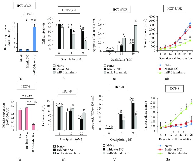
Although aberrant expression of miR-34a, an essential tumor suppressor miRNA, has been frequently observed in colon cancer (CCa), whether miR-34a can regulate CCa progression by modulating other facets of this malignancy (such as multidrug resistance, MDR) remains unknown. Here, we report for the first time that miR-34a expression was significantly downregulated in clinical CCa samples from oxaliplatin-resistant patients and in experimentally established multidrug-resistant CCa cells. By using histoculture drug response assay, we further confirmed that clinical CCa samples with lower miR-34a expression appeared to be more resistant to chemotherapy. Functionally, ectopic expression of exogenous miR-34a resensitized multidrug-resistant HCT-8/OR cells to oxaliplatin treatment, whereas miR-34a inhibition augmented the oxaliplatin resistance in chemosensitive HCT-8 cells. Mechanistically, miR-34a positively regulated the mRNA stability of the ornithine decarboxylase antizyme 2 (OAZ2) by directly targeting its three prime untranslated region (3'UTR). Consequently, suppression of the expression of miR-34a/OAZ2 signaling by chemotherapeutic agents significantly enhanced the activation of MDR-associated ATP-binding cassette (ABC) transporters and antiapoptosis pathways, thus leading to MDR development in CCa cells. Collectively, our combined analysis reveals a critical role of miR-34a/OAZ2 cascade in conferring a proper cellular response to CCa chemotherapy.


Figures

Figure 1
Figure 1
Downregulation of miR-34a expression is associated with multidrug resistance in colon cancer (CCa). (a) miR-34a expression in normal colon tissues, primary CCa tissues, and recurrent CCa tissues was determined using RT-qPCR. Fold change was determined for each sample relative to the internal control gene 18S. Each value is a mean ± S.E.M. from three experiments. (b) CCa cells (2.0 × 104/well) were seeded in a 96-well plate. 24 h later, cells were treated with different concentrations of oxaliplatin, paclitaxel, and vincristine as indicated, for another 48 h. Subsequent cell viability was determined using the WST-1 viability assay. The IC50 values (drug concentrations that achieved 50% growth inhibition) were determined as described in Materials and Methods. (c) Characterization of miR-34a expression in different CCa cells using RT-qPCR. (d) Histoculture drug response assay (HDRA) showing the differential chemosensitivities in clinical CCa tissues with different levels of miR-34a expression. P < 0.05 and ∗∗ P < 0.01 when compared to the Naive cells.
Figure 2
Figure 2
Manipulation of miR-34a expression affects chemosensitivity in CCa cells. (a) HCT-8/OR cells were transfected with miR-34a mimic or Mimic negative control (NC) as described in Materials and Methods. 48 h later, cells were collected, and the relative expression levels of miR-34a were assayed using RT-qPCR. HCT-8/OR cells with different transfections were incubated with different doses of oxaliplatin for 24 h, followed by cell viability assay (b) and apoptosis evaluation (c). Different superscript letters denote groups that are statistically different (P < 0.05). (d) HCT-8/OR cell-derived tumor xenograft model was established as described in Materials and Methods. Tumor volumes were measured every 4 days. P < 0.05 and ∗∗ P < 0.01 when comparing miR-34a mimic to Mimic NC. (e) HCT-8 cells were transfected with miR-34a inhibitor or Inhibitor NC as described in Materials and Methods. 48 h later, cells were collected and the relative expression levels of miR-34a were assayed using RT-qPCR. HCT-8 cells with different transfections were incubated with different doses of oxaliplatin for 24 h, followed by cell viability assay (f) and apoptosis evaluation (g). Different superscript letters denote groups that are statistically different (P < 0.05). (h) HCT-8 cell-derived tumor xenograft model was established as described in Materials and Methods. Tumor volumes were measured every 4 days. P < 0.05 and ∗∗ P < 0.01 when comparing miR-34a inhibitor to Inhibitor NC.
Figure 3
Figure 3
miR-34a inhibits the expression levels of signaling pathways associated with multidrug resistance. (a) Immunoblotting analysis of the expression levels of MRP2, P-gp, BCRP, and Bcl-2 in HCT-8 and HCT-8/OR cells. β-Actin served as loading control. (b) 48 h after transfection with miR-34a mimic or Mimic NC, HCT-8/OR cells were subjected to immunoblotting analysis of the expression levels of MRP2, P-gp, BCRP, and Bcl-2. (c) 48 h after transfection with miR-34a inhibitor or Inhibitor NC, HCT-8 cells were subjected to immunoblotting analysis of the expression levels of MRP2, P-gp, BCRP, and Bcl-2. (d) Immunoblotting analysis of the expression levels of MRP2, P-gp, BCRP, and Bcl-2 in HCT-116 and HCT-116/OR cells. (e) 48 h after transfection with miR-34a mimic or Mimic NC, HCT-116/OR cells were subjected to immunoblotting analysis of the expression levels of MRP2, P-gp, BCRP, and Bcl-2. (f) 48 h after transfection with miR-34a inhibitor or Inhibitor NC, HCT-116 cells were subjected to immunoblotting analysis of the expression levels of MRP2, P-gp, BCRP, and Bcl-2. (g) Immunoblotting analysis of the expression levels of MRP2, P-gp, BCRP, and Bcl-2 in SW-480 and SW-480/OR cells. (h) 48 h after transfection with miR-34a mimic or Mimic NC, SW-480/OR cells were subjected to immunoblotting analysis of the expression levels of MRP2, P-gp, BCRP, and Bcl-2. (i) 48 h after transfection with miR-34a inhibitor or Inhibitor NC, SW-480 cells were subjected to immunoblotting analysis of the expression levels of MRP2, P-gp, BCRP, and Bcl-2.
Figure 4
Figure 4
Direct regulation of OAZ2 expression by miR-34a. (a) The binding site of miR-34a in the OAZ2 3′UTR was analyzed using miRanda database. (b) Relative expression levels of miR-34a and OAZ2 mRNA in clinical CCa samples were evaluated using RT-qPCR, followed by Pearson chi-square test. (c) Immunoblotting analysis of OAZ2 expression in HCT-8 and HCT-8/OR cells with different transfections. (d) RT-qPCR analysis of OAZ2 mRNA expression in HCT-8 and HCT-8/OR cells with different transfections. (e) Simplified structure of the potential binding site of miR-34a onto the wild-type (WT) or mutant (Mu) OAZ2 3′UTR. (f) 0.5 μg of pGL3-OAZ2 WT or pGL3-OAZ2 Mu, along with 0.05 μg of pRL-SV40 plasmid and miR-34a mimic, was cotransfected into HeLa cells. 48 h later, cells were harvested and subjected to luciferase activity measurements.
Figure 5
Figure 5
OAZ2 overexpression abolishes miR-34a deficiency-induced oxaliplatin-resistance. (a) HCT-8/OAZ2 cells were established as described in Materials and Methods. OAZ2 overexpression was confirmed by immunoblotting analysis. pcDNA3.1+/DYK vector is expressed in mammalian cells as a tagged protein with a C-terminal DYKDDDDK tag (DYKDDDDK is the same as FLAG® which is a registered trademark of Sigma-Aldrich). (b) RT-qPCR analysis of miR-34a expression in HCT-8/OAZ2 cells. (c) HCT-8/OAZ2 cells with different transfections were incubated with different doses of oxaliplatin for 24 h, followed by cell viability assay. Different superscript letters denote groups that are statistically different (P < 0.05). (d) HCT-8/OAZ2 cell-derived tumor xenografts model was established as described in Materials and Methods. Tumor volumes were measured every 4 days. P < 0.05 and ∗∗ P < 0.01 when comparing DYK-OAZ2 + miR-34a inhibitor to miR-34a inhibitor alone. (e) HCT-8/OAZ2 cells were transiently transfected with miR-34a inhibitor or Inhibitor NC for 48 h, followed by immunoblotting analysis of the expression levels of OAZ2, MRP2, P-gp, BCRP, and Bcl-2. (f) Proposed working model in the current study.

Similar articles

Cited by

References

    1. Thomas J., Ohtsuka M., Pichler M., Ling H. MicroRNAs: clinical relevance in colorectal cancer. International Journal of Molecular Sciences. 2015;16(12):28063–28076. doi: 10.3390/ijms161226080. - DOI - PMC - PubMed
    1. Liu Y. R., Liang L., Zhao J. M., et al. Twist1 confers multidrug resistance in colon cancer through upregulation of ATP-binding cassette transporters. Oncotarget. 2017;8(32):52901–52912. doi: 10.18632/oncotarget.17548. - DOI - PMC - PubMed
    1. Calcagno A. M., Fostel J. M., To K. K. W., et al. Single-step doxorubicin-selected cancer cells overexpress the ABCG2 drug transporter through epigenetic changes. British Journal of Cancer. 2008;98(9):1515–1524. doi: 10.1038/sj.bjc.6604334. - DOI - PMC - PubMed
    1. Lubelska K., Wiktorska K., Mielczarek L., Milczarek M., Zbroinska-Bregisz I., Chilmonczyk Z. Sulforaphane regulates NFE2L2/Nrf2-dependent xenobiotic metabolism phase II and phase III enzymes differently in human colorectal cancer and untransformed epithelial colon cells. Nutrition and Cancer. 2016;68(8):1338–1348. doi: 10.1080/01635581.2016.1224369. - DOI - PubMed
    1. Ebert B., Kisiela M., Wsol V., Maser E. Proteasome inhibitors MG-132 and bortezomib induce AKR1C1, AKR1C3, AKR1B1, and AKR1B10 in human colon cancer cell lines SW-480 and HT-29. Chemico-Biological Interactions. 2011;191(1-3):239–249. doi: 10.1016/j.cbi.2010.12.026. - DOI - PubMed

MeSH terms

LinkOut - more resources