Review
. 2018 Jul;6(4):250-259.

Meniscal Root Tears: Current Concepts Review

Affiliations
Review

Meniscal Root Tears: Current Concepts Review

Santiago Pache et al. Arch Bone Jt Surg. 2018 Jul.

Abstract

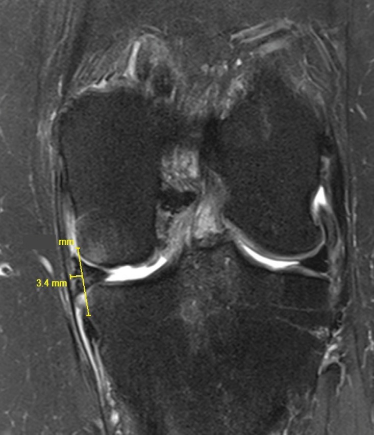
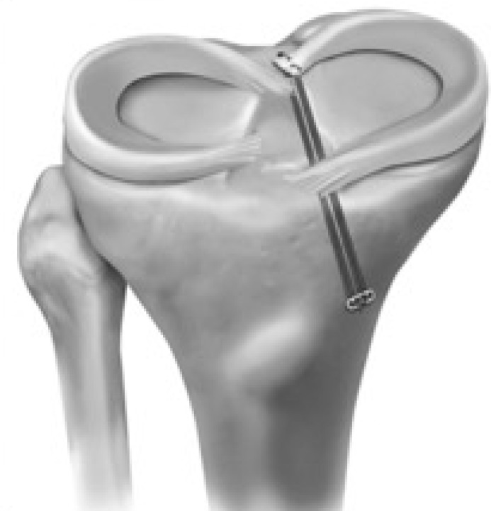
Meniscal root tears are defined as radial tears located within 1 cm from the meniscal attachment or a bony root avulsion. This injury is biomechanically comparable to a total meniscectomy, leading to compromised hoop stresses resulting in decreased tibiofemoral contact area and increased contact pressures in the involved compartment. These changes are detrimental to the articular cartilage and ultimately lead to the development of early osteoarthritis. Surgical repair is the treatment of choice in patients without significant osteoarthritis (Outerbridge grades 3 or 4). Root repairs have been reported to improve clinical outcomes, decrease meniscal extrusion and slow the onset of degenerative changes. Here we describe the anatomy, biomechanics, clinical evaluation, treatment methods, outcomes, and post-operative rehabilitation for posterior meniscal root tears.

Keywords: Lateral meniscus; Medial meniscus; Meniscus root tear; Outcomes; Root repair.


Figures

Figure 1
Figure 1
Medial and lateral meniscal posterior root attachments and relevant arthroscopic bony landmarks. (A) Superior view and (B) posterior view. ACL, anterior cruciate ligament; LPRA, lateral meniscus posterior root attachment; LTE, lateral tibial eminence; MPRA, medial meniscus posterior root attachment; MTE, medial tibial eminence; PCL, posterior cruciate ligament bundle attachments; SWF, shiny white fibers of posterior horn of medial meniscus. (Reproduced with permission from: Johannsen M, Civitarese DM, Padalecki JR, Goldsmith MT, Wijdicks CA, LaPrade RF. Qualitative and quantitative anatomic analysis of the posterior root attachments of the medial and lateral menisci. Am J Sport Med. 2012;40(10):2342–7).
Figure 2
Figure 2
The anterior lateral (AL) meniscal root is shown to run deeply beneath and overlap with the anterior cruciate ligament (ACL), as observed in all knees. The anterior medial (AM) meniscal root is depicted with supplemental fibers (SFs) that were observed to be anterior and distal to the central root. Anterior meniscal root structure is also described in relation to pertinent bony and soft tissue landmarks. AC, articular cartilage; LTE, lateral tibial eminence; MTE, medial tibial eminence; PL root, posterior lateral meniscal root; PM root, posterior medial meniscal root; TT, tibial tuberosity. (Reproduced with permission from: LaPrade CM, Ellman MB, Rasmussen MT, James EW, Wijdicks CA, Engebretsen L, et al. Anatomy of the anterior root attachments of the medial and lateral menisci: a quantitative analysis. Am J Sport Med. 2014;42(10):2386–92.)
Figure 3
Figure 3
Meniscal root tear classification system illustrated in posterior medial meniscal root. Type 1: partial stable tear. Type 2: Complete radial tear within 9 mm from attachment. Type 3: Bucket-handle tear with complete root detachment. Type 4: Complex oblique or longitudinal tear with complete root detachment. Type 5: Bony avulsion fracture of the root attachment. (Reproduced with permission from: LaPrade CM, James EW, Cram TR, Feagin JA, Engebretsen L, LaPrade RF. Meniscal Root Tears. Am J Sports Med. 2015;43(2):363-9
Figure 4
Figure 4
Left knee MRI showing a medial meniscus posterior root tear (MPRT) (White arrow). A: Sagittal plane view. Ghost sign; B: Axial plane view; C: Coronal plane view
Figure 5
Figure 5
Left knee MRI showing a medial meniscus extrusion on the coronal plane views.
Figure 6
Figure 6
Transtibial pull-out repair of a medial meniscal posterior root tear in a right knee. (Reproduced with permission from: LaPrade CM, LaPrade MD, Turnbull TL, Wijdicks CA, LaPrade RF. Biomechanical evaluation of the transtibial pull-out technique for posterior medial meniscal root repairs using 1 and 2 transtibial bone tunnels. Am J Sports Med. 2015;43(4):899–904).

References

    1. Choi CJ, Choi YJ, Lee JJ, Choi CH. Magnetic resonance imaging evidence of meniscal extrusion in medial meniscus posterior root tear. Arthroscopy. 2010;26(12):1602–6. - PubMed
    1. Laprade CM, Jansson KS, Dornan G, Smith SD, Wijdicks CA, Laprade RF. Altered tibiofemoral contact mechanics due to lateral meniscus posterior horn root avulsions and radial tears can be restored with in situ pull-out suture repairs. J Bone Joint Surg Am. 2014;96(6):471–9. - PubMed
    1. Padalecki JR, Jansson KS, Smith SD, Dornan GJ, Pierce CM, Wijdicks CA, et al. Biomechanical consequences of a complete radial tear adjacent to the medial meniscus posterior root attachment site: in situ pull-out repair restores derangement of joint mechanics. Am J Sports Med. 2014;42(3):699–707. - PubMed
    1. Marzo JM, Gurske-DePerio J. Effects of medial meniscus posterior horn avulsion and repair on tibiofemoral contact area and peak contact pressure with clinical implications. Am J Sports Med. 2009;37(1):124–9. - PubMed
    1. Koenig JH, Ranawat AS, Umans HR, Difelice GS. Meniscal root tears: diagnosis and treatment. Arthroscopy. 2009;25(9):1025–32. - PubMed

LinkOut - more resources