

Utility of Optical Density of Picrosirius Red Birefringence for Analysis of Cross-Linked Collagen in Remodeling of the Peripartum Cervix for Parturition
- PMID: 30175325
- PMCID: PMC6117116
- DOI: 10.31038/IGOJ.2018121
Utility of Optical Density of Picrosirius Red Birefringence for Analysis of Cross-Linked Collagen in Remodeling of the Peripartum Cervix for Parturition
Abstract
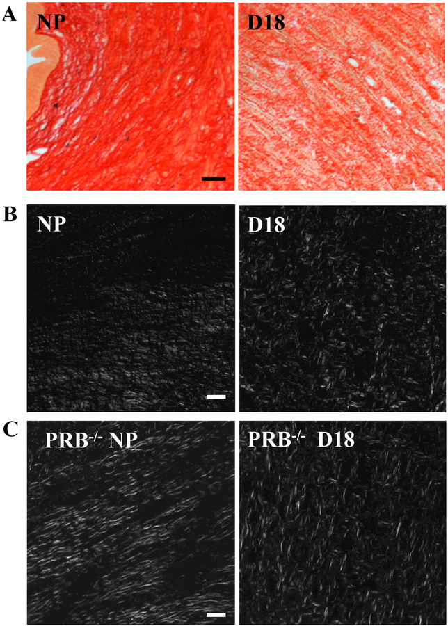
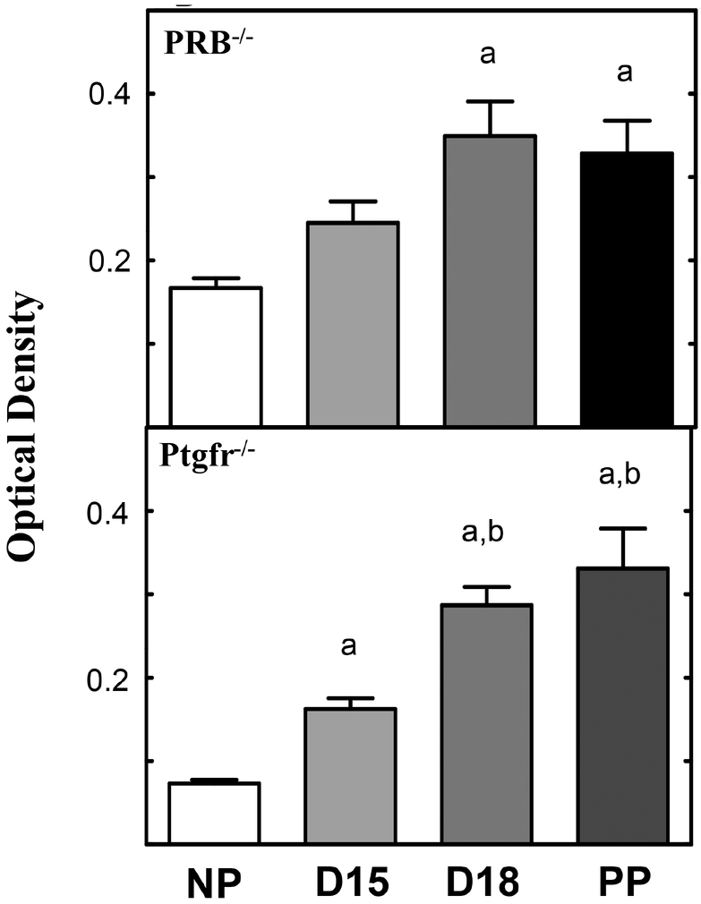
We report on development of a rapid, quantitative analysis technique of collagen fibers in cross-linked structures to assess remodeling of the cervix during the transition from soft to ripening in preparation for birth. Optical density analysis of picrosirius red stain tissue using circular polarized birefringence light from fixed paraffin-embedded or frozen cervix from pregnant mice during phases of remodeling prior to birth. Data were analyzed using NIH Image J and extended recently to include studies of prepartum cervix in peripartum women. Our results, developed a rapid, consistent, technique to quantify cervical organization. This approach assesses the structure of collagen organization (the principle component of the cervix) and is essential for analysis of experimental outcomes that disrupt cervical morphology in rodent models of preterm birth. The technique, in this report has, for the first time permitted rapid, accurate assessment of the stages that define cervical ripening with large numbers of slides from individual animals. The approach integrates analysis of collagen organization, with distensability and inflammation, processes associated with cervical change before birth. This analysis further holds promise to evaluate other tissues, but also fibrolytic and fibrogenic changes in collagen associated with physiological or pathophysiological conditions.
Keywords: inflammation; pregnancy; remodeling; ripening.
Conflict of interest statement
Competing Interests All authors declare and affirm the lack of competing interests, finical or otherwise, in this manuscript.
Figures
Similar articles
-
Increased innervation and ripening of the prepartum murine cervix.J Soc Gynecol Investig. 2005 Dec;12(8):578-85. doi: 10.1016/j.jsgi.2005.08.006. J Soc Gynecol Investig. 2005. PMID: 16325747
-
Immunobiology of Cervix Ripening.Front Immunol. 2020 Jan 24;10:3156. doi: 10.3389/fimmu.2019.03156. eCollection 2019. Front Immunol. 2020. PMID: 32038651 Free PMC article. Review.
-
Contributions to the dynamics of cervix remodeling prior to term and preterm birth.Biol Reprod. 2017 Jan 1;96(1):13-23. doi: 10.1095/biolreprod.116.142844. Biol Reprod. 2017. PMID: 28395330 Free PMC article. Review.
-
Density of Stromal Cells and Macrophages Associated With Collagen Remodeling in the Human Cervix in Preterm and Term Birth.Reprod Sci. 2016 May;23(5):595-603. doi: 10.1177/1933719115616497. Epub 2015 Nov 25. Reprod Sci. 2016. PMID: 26608218 Free PMC article.
-
Progesterone regulation of cervix ripening in preterm and term birth.bioRxiv [Preprint]. 2025 Feb 1:2025.01.31.636012. doi: 10.1101/2025.01.31.636012. bioRxiv. 2025. PMID: 39974958 Free PMC article. Preprint.
Cited by
-
Photoacoustic imaging of the uterine cervix to assess collagen and water content changes in murine pregnancy.Biomed Opt Express. 2019 Aug 19;10(9):4643-4655. doi: 10.1364/BOE.10.004643. eCollection 2019 Sep 1. Biomed Opt Express. 2019. PMID: 31565515 Free PMC article.
-
Mechanics of cervical remodelling: insights from rodent models of pregnancy.Interface Focus. 2019 Oct 6;9(5):20190026. doi: 10.1098/rsfs.2019.0026. Epub 2019 Aug 16. Interface Focus. 2019. PMID: 31485313 Free PMC article. Review.
-
Label-free optical imaging of cell function and collagen structure for cell-based therapies.Curr Opin Biomed Eng. 2023 Mar;25:100433. doi: 10.1016/j.cobme.2022.100433. Epub 2022 Dec 9. Curr Opin Biomed Eng. 2023. PMID: 36642995 Free PMC article.
-
Collagen molecular organization preservation in human fascia lata and periosteum after tissue engineering.Front Bioeng Biotechnol. 2024 Apr 3;12:1275709. doi: 10.3389/fbioe.2024.1275709. eCollection 2024. Front Bioeng Biotechnol. 2024. PMID: 38633664 Free PMC article.
-
N,N-dimethylacetamide blocks inflammation-induced preterm birth and remediates maternal systemic immune responses.bioRxiv [Preprint]. 2025 Jan 20:2025.01.16.633350. doi: 10.1101/2025.01.16.633350. bioRxiv. 2025. Update in: Sci Rep. 2025 Mar 10;15(1):8234. doi: 10.1038/s41598-025-93282-0. PMID: 39896567 Free PMC article. Updated. Preprint.
References
-
- Bracegirdle B (1977) The History of Histology: A Brief Survey of Sources. History of Science 15: 77–101.
-
- Mescher AL, Junqueira LCU (2016) Junqueira’s basic histology: Text and atlas (Fourteenth edition). New York: McGraw-Hill Education.
-
- Sabiston DC, Townsend CM (2012) Sabiston Textbook of Surgery: The Biological Basis of Modern Surgical Practice. Elsevier Saunders.
-
- Junqueira LC, Zugaib M, Montes GS, Toledo OM, Krisztan RM, Shigihara KM (1980) Morphologic and histochemical evidence for the occurrence of collagenolysis and for the role of neutrophilic polymorphonuclear leukocytes during cervical dilation. Am J Obstet Gynecol. 138: 273–81. - PubMed
-
- Uldbjerg N, Ekman G, Malmstrom A, Olsson K, Ulmsten U (1983) Ripening of the human uterine cervix related to changes in collagen, glycosaminoglycans, and collagenolytic activity. Am J Obstet Gynecol 147: 662–6. - PubMed
Grants and funding
LinkOut - more resources
Full Text Sources