

Feedforward- and motor effort-dependent increase in prefrontal oxygenation during voluntary one-armed cranking
- PMID: 30175404
- PMCID: PMC6209741
- DOI: 10.1113/JP276956
Feedforward- and motor effort-dependent increase in prefrontal oxygenation during voluntary one-armed cranking
Abstract
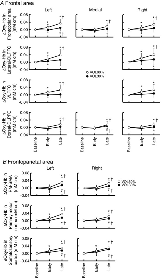
Key points: Some cortical areas are believed to transmit a descending signal in association with motor intention and/or effort that regulates the cardiovascular system during exercise (termed central command). However, there was no evidence for the specific cortical area responding prior to arbitrary motor execution and in proportion to the motor effort. Using a multichannel near-infrared spectroscopy system, we found that the oxygenation of the dorsolateral and ventrolateral prefrontal cortices on the right side increases in a feedforward- and motor effort-dependent manner during voluntary one-armed cranking with the right arm. This finding may suggest a role of the dorsolateral and ventrolateral prefrontal cortices in triggering off central command and may help us to understand impaired regulation of the cardiovascular system in association with lesion of the prefrontal cortex.
Abstract: Output from higher brain centres (termed central command) regulates the cardiovascular system during exercise in a feedforward- and motor effort-dependent manner. This study aimed to determine a cortical area responding prior to arbitrarily started exercise and in proportion to the effort during exercise. The oxygenation responses in the frontal and frontoparietal areas during one-armed cranking with the right arm were measured using multichannel near-infrared spectroscopy, as indexes of regional blood flow responses, in 20 subjects. The intensity of voluntary exercise was 30% and 60% of the maximal voluntary effort (MVE). At the start period of both voluntary cranking tasks, the oxygenation increased (P < 0.05) only in the lateral and dorsal part of the dorsolateral prefrontal cortex (DLPFC), ventrolateral prefrontal cortex (VLPFC) and sensorimotor cortices. Then, the oxygenation increased gradually in all cortical areas during cranking at 60% MVE, while oxygenation increased only in the frontoparietal area and some of the frontal area during cranking at 30% MVE. The rating of perceived exertion to the cranking tasks correlated (P < 0.05) with the oxygenation responses on the right side of the lateral-DLPFC (r = 0.46) and VLPFC (r = 0.48) and the frontopolar areas (r = 0.47-0.49). Motor-driven passive one-armed cranking decreased the oxygenation in most cortical areas, except the contralateral frontoparietal areas. Accordingly, the lateral-DLPFC and VLPFC on the right side would respond in a feedforward- and motor effort-dependent manner during voluntary exercise with the right arm. Afferent inputs from mechanosensitive afferents may decrease the cortical oxygenation.
Keywords: central command; dorsolateral and ventrolateral prefrontal cortices; motor effort.
© 2018 The Authors. The Journal of Physiology © 2018 The Physiological Society.
Figures


Similar articles
-
Central command increases muscular oxygenation of the non-exercising arm at the early period of voluntary one-armed cranking.Physiol Rep. 2017 Apr;5(7):e13237. doi: 10.14814/phy2.13237. Physiol Rep. 2017. PMID: 28381448 Free PMC article.
-
An increase in prefrontal oxygenation at the start of voluntary cycling exercise was observed independently of exercise effort and muscle mass.Eur J Appl Physiol. 2018 Aug;118(8):1689-1702. doi: 10.1007/s00421-018-3901-4. Epub 2018 May 31. Eur J Appl Physiol. 2018. PMID: 29855789
-
Increased oxygenation of the cerebral prefrontal cortex prior to the onset of voluntary exercise in humans.J Appl Physiol (1985). 2015 Sep 1;119(5):452-62. doi: 10.1152/japplphysiol.00406.2015. Epub 2015 Jul 16. J Appl Physiol (1985). 2015. PMID: 26183481
-
Connections underlying the synthesis of cognition, memory, and emotion in primate prefrontal cortices.Brain Res Bull. 2000 Jul 15;52(5):319-30. doi: 10.1016/s0361-9230(99)00245-2. Brain Res Bull. 2000. PMID: 10922509 Review.
-
Cognitive control and right ventrolateral prefrontal cortex: reflexive reorienting, motor inhibition, and action updating.Ann N Y Acad Sci. 2011 Apr;1224(1):40-62. doi: 10.1111/j.1749-6632.2011.05958.x. Ann N Y Acad Sci. 2011. PMID: 21486295 Free PMC article. Review.
Cited by
-
Central command suppresses pressor-evoked bradycardia at the onset of voluntary standing up in conscious cats.Exp Physiol. 2023 Jan;108(1):28-37. doi: 10.1113/EP090718. Epub 2022 Nov 20. Exp Physiol. 2023. PMID: 36404613 Free PMC article.
-
Physiological role of anticipatory cardiorespiratory responses to exercise.Physiol Rep. 2022 Mar;10(5):e15210. doi: 10.14814/phy2.15210. Physiol Rep. 2022. PMID: 35246949 Free PMC article.
-
Effect of Combined Mental Task and Metaboreflex Activation on Hemodynamics and Cerebral Oxygenation in Patients With Metabolic Syndrome.Front Physiol. 2020 May 14;11:397. doi: 10.3389/fphys.2020.00397. eCollection 2020. Front Physiol. 2020. PMID: 32477157 Free PMC article.
-
Cerebral Oxygenation Dynamics During Incremental Exercise: Comparison of Arm Cranking and Leg Cycling.Adv Exp Med Biol. 2021;1269:125-130. doi: 10.1007/978-3-030-48238-1_20. Adv Exp Med Biol. 2021. PMID: 33966206
-
Data Processing in Functional Near-Infrared Spectroscopy (fNIRS) Motor Control Research.Brain Sci. 2021 May 9;11(5):606. doi: 10.3390/brainsci11050606. Brain Sci. 2021. PMID: 34065136 Free PMC article. Review.
References
-
- Arnsten AF (1997). Catecholamine regulation of the prefrontal cortex. J Psychopharmacol 11, 151–162. - PubMed
-
- Asahara R, Endo K, Liang N & Matsukawa K (2018). An increase in prefrontal oxygenation at the start of voluntary cycling exercise was observed independently of exercise effort and muscle mass. Eur J Appl Physiol 118, 1689–1702. - PubMed
-
- Asahara R & Matsukawa K (2018). Decreased prefrontal oxygenation elicited by stimulation of limb mechanosensitive afferents during cycling exercise. Am J Physiol Regul Integr Comp Physiol 315, R230–R240. - PubMed
-
- Asahara R, Matsukawa K, Ishii K, Liang N & Endo K (2016). The prefrontal oxygenation and ventilatory responses at start of one‐legged cycling exercise have relation to central command. J Appl Physiol 121, 1115–1126. - PubMed
-
- Bernard RA, Goran DA, Sakai ST, Carr TH, McFarlane D, Nordell B, Cooper TG & Potchen EJ (2002). Cortical activation during rhythmic hand movements performed under three types of control: an fMRI study. Cogn Affect Behav Neurosci 2, 271–281. - PubMed
Publication types
MeSH terms
Grants and funding
LinkOut - more resources
Full Text Sources
Other Literature Sources