. 2018 Sep 3;37(1):210.
doi: 10.1186/s13046-018-0889-x.

Corosolic acid, a natural triterpenoid, induces ER stress-dependent apoptosis in human castration resistant prostate cancer cells via activation of IRE-1/JNK, PERK/CHOP and TRIB3

Affiliations

Corosolic acid, a natural triterpenoid, induces ER stress-dependent apoptosis in human castration resistant prostate cancer cells via activation of IRE-1/JNK, PERK/CHOP and TRIB3

Bo Ma et al. J Exp Clin Cancer Res. .

Abstract

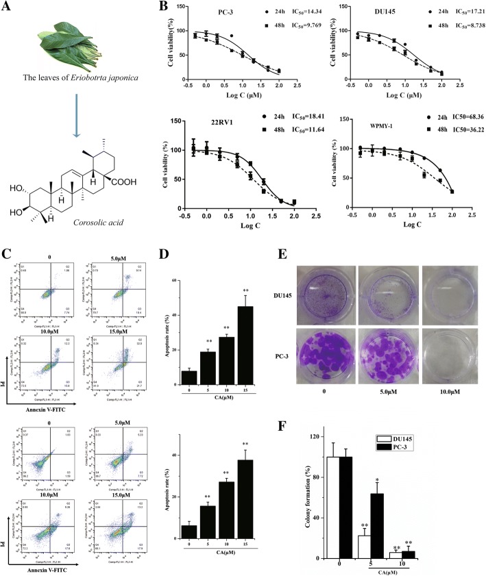
Background: The development of potent non-toxic chemotherapeutic drugs against castration resistant prostate cancer (CRPC) remains a major challenge. Corosolic acid (CA), a natural triterpenoid, has anti-cancer activity with limited side effects. However, CA anti-prostate cancer activities and mechanisms, particularly in CRPC, are not clearly understood. In this study, we investigated CA anti-tumor ability against human CRPC and its mechanism of action.

Methods: The cell apoptosis and proliferation effects were evaluated via MTT detection, colony formation assay and flow cytometry. Western blot, gene transfection and immunofluorescence assay were applied to investigate related protein expression of Endoplasmic reticulum stress. A xenograft tumor model was established to investigate the inhibitory effect of CA on castration resistant prostate cancer in vivo.

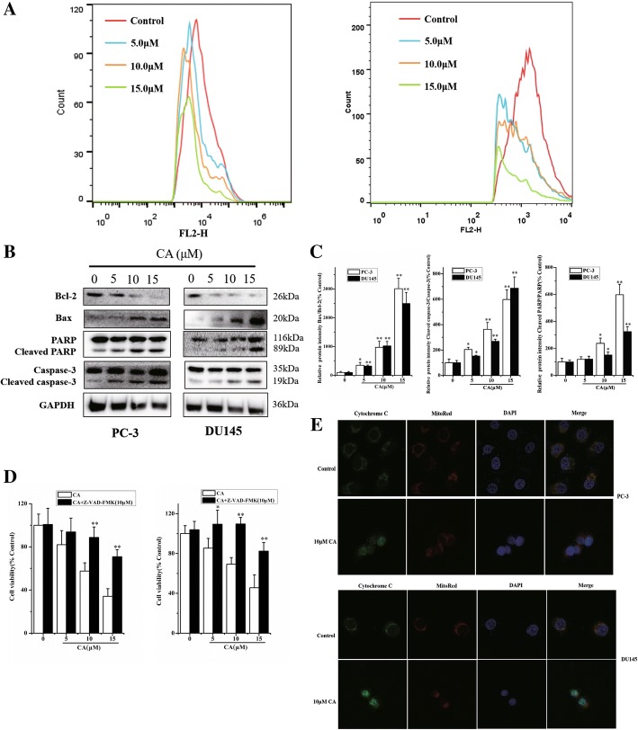
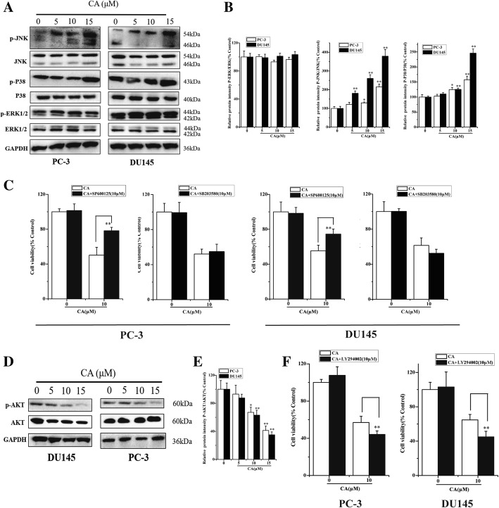
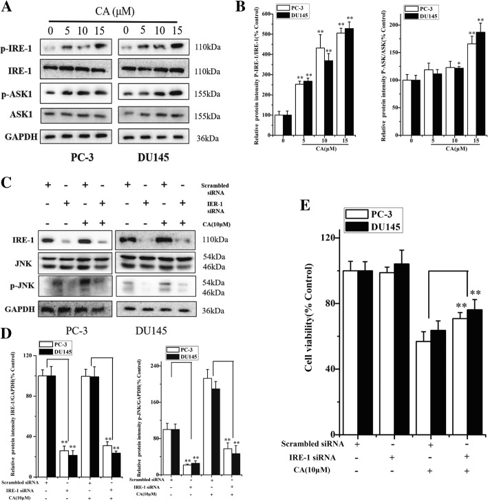
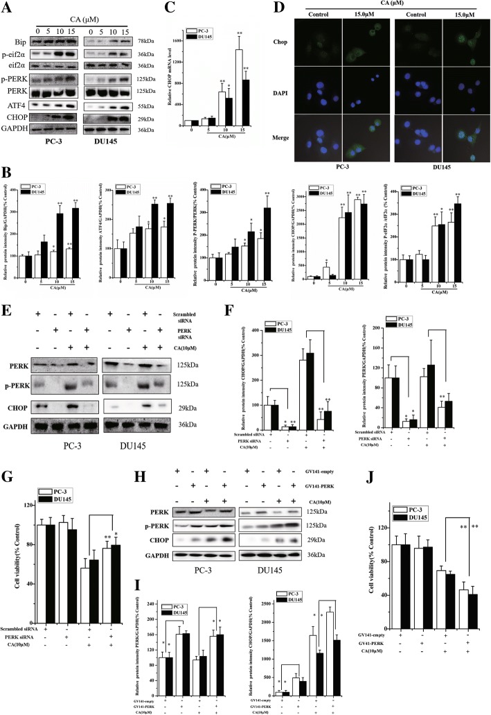
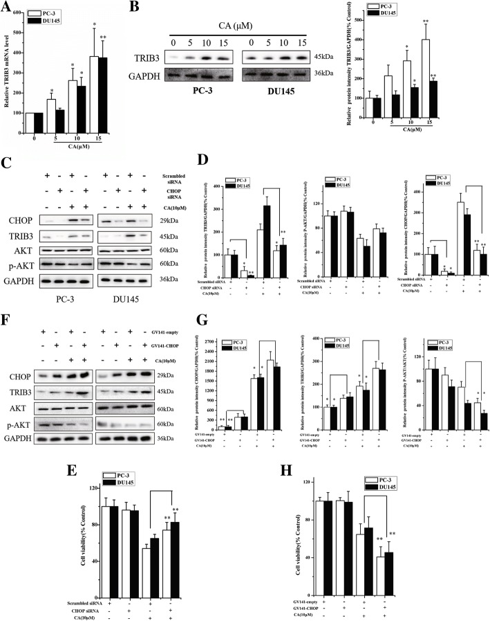
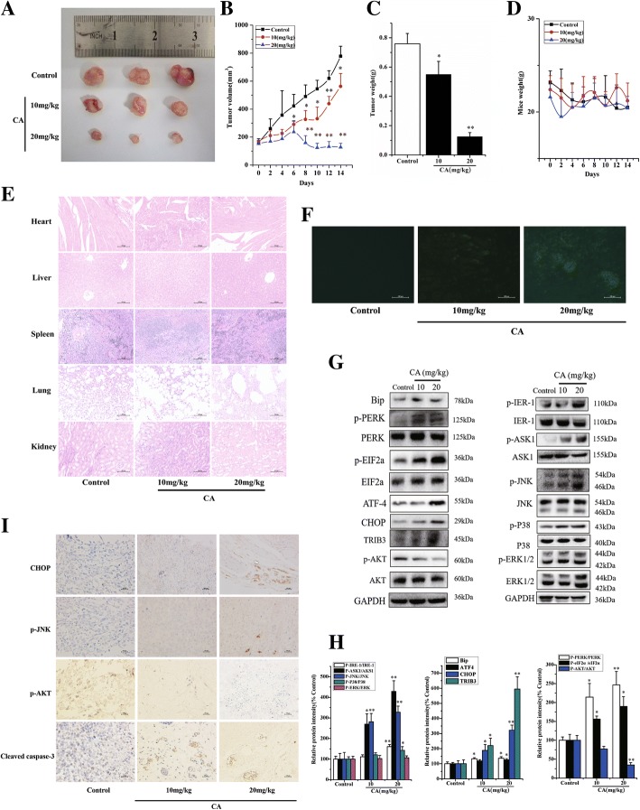
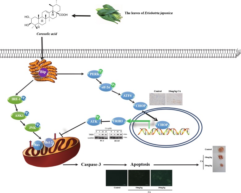
Results: The results showed that CA inhibited cell growth and induced apoptosis in human prostate cancer cell (PCa) line PC-3 and DU145, as well as retarded tumor growth in a xenograft model, exerting a limited toxicity to normal cells and tissues. Importantly, CA activated endoplasmic reticulum (ER) stress-associated two pro-apoptotic signaling pathways, as evidenced by increased protein levels of typical ER stress markers including IRE-1/ASK1/JNK and PERK/eIF2α/ATF4/CHOP. IRE-1, PERK or CHOP knockdown partially attenuated CA cytotoxicity against PCa cells. Meanwhile, CHOP induced expression increased Tribbles 3 (TRIB3) level, which lead to AKT inactivation and PCa cell death. CHOP silencing resulted in PCa cells sensitive to CA-induced apoptosis.

Conclusion: Our data demonstrated, for the first time, that CA might represent a novel drug candidate for the development of an anti-CRPC therapy.

Keywords: CCAAT-enhancer-binding protein homologous protein (CHOP); Castration resistant PCa (CRPC); Corosolic acid (CA); Endoplasmic reticulum stress (ER stress); Protein kinase RNA-like endoplasmic reticulum kinase (PERK); Tribbles homolog 3 (TRIB3).


Conflict of interest statement

Ethics approval and consent to participate

For the animal study, all procedures performed on the animals in this study were approved (NO. NJTECH-AE-2017006) the Guidelines for the Animal Ethics Committee of Nanjing Tech University.

Consent for publication

Not applicable.

Competing interests

The authors declare that they have no competing interests.

Publisher’s Note

Springer Nature remains neutral with regard to jurisdictional claims in published maps and institutional affiliations.

Figures

Fig. 1
Fig. 1
CA suppresses cell proliferation and induces apoptosis in human prostate cancer cells. a Structure of the CA molecule. b PC-3, DU145, 22rv1 and WPMY-1 cells were treated with various concentrations of CA for 24 and 48 h, cell viability was assayed by MTT assay. c Apoptosis index in PC-3 and DU145 cells with 0, 5, 10 and 15 μM CA treatments for 24 h detected by annexinV/PI flow cytometry assay. d Statistical analysis result of flow cytometric analysis of apoptosis (both of early and later apoptosis). e Cell proliferation was measured by colony formation in 12-well plates with crystal violet staining. Representative photographs are shown. f The percentage of colony formation was calculated by defining the number of colonies in the absence of CA as 100%. The results are presented as mean ± SD and described as column chart *p < 0.05 and **p < 0.01 as compared with untreated control
Fig. 2
Fig. 2
Effect of CA on mitochondria dependent apoptosis in human prostate cancer cells. a Mitochondrial membrane potentials (ΔΨm) were assessed with JC-1 staining by flow cytometry. b The expression of cell key apoptosis-related proteins was detected by Western-blotting and they were statistically analyzed (c). The PC-3 and DU145 cells was pre-treated with caspase inhibitor (10 μM Z-VAD-FMK) for 3 h and followed by incubation with or without CA for 24 h, respectively. d The cell viability was determined by MTT assay. The results are presented as mean ± SD and described as column chart *p < 0.05 and **p < 0.01 as compared with corresponding group. e Effect of CA induced-Cytochrome c release was analyzed by immunofluorescence staining
Fig. 3
Fig. 3
MAPK and AKT signaling pathways are involved in CA apoptotic effect on human PCa cells. MAPKs (a) and AKT (d) protein expressions were analyzed using western blot. Densitometry scanning analysis for ratio of p-JNK/total JNK, p-P38/total P38 and p-ERK1/2/total ERK1/2 (b) as well as p-AKT/total AKT (e). Data represent the mean ± SD of three independent experiments. *p < 0.05 and **p < 0.01 as compared with untreated control. c Effects of SP600125, SB203580 and LY294002 (f) CA-induced apoptosis in cultured cells for 24 h. Cell viability was determined by MTT assay and data represent the mean ± SD of six experiments in each group. *p < 0.05, **p < 0.01 as compared with single CA treated cells
Fig. 4
Fig. 4
IRE-1-mediated ASK1-JNK activation plays a key role in CA-induced cell death. aThe PC-3 and DU145 cells were treated, respectively, by CA for 24 h at doses of 5, 10, 15 μM. The p-IRE-1, IRE-1, p-ASK1 and ASK1 protein expression were measured by Western blotting, and the bands were scanned and data of statistically analysis were listed in (b). Data represent the mean ± SD of three independent experiments. *p < 0.05 and **p < 0.01 as compared with untreated control. The cells transfected with IRE-1 siRNA or control siRNA were treated with CA (10 μM) for 24 h. The protein expression was measured by western blotting (c), Densitometric analysis of protein bands was showed in (d). eThe cell viability was determined by MTT assay. The results are presented as mean ± SD and represent three individual experiments. *p < 0.05 and **p < 0.01 as compared with corresponding group
Fig. 5
Fig. 5
CA activates the PERK-eIF2a-ATF4 pathway leading to the pro-apoptotic CHOP up-regulation in human PCa cells. The PC-3 and DU145 cells were treated with CA at the indicated concentrations (5, 10, 15 μM.) for 24 h. a the phosphorylation of PERK, eIF2α and expression levels of Bip, PERK, eIF2α, ATF4 and CHOP were measured by western blot using corresponding antibodies. b And Protein levels were quantified by grayscale scan and the GAPDH was used as the loading control. c Total RNAs were extracted and subjected to real-time PCR analysis using specific primer sets for CHOP and GAPDH, and the data were normalized to GAPDH expression. The results are presented as mean ± SD and represent three individual experiments. *p < 0.05 and **p < 0.01 as compared with untreated control. d Effects of CA induced-CHOP activation analyzed by immunofluorescence staining. The PC-3 and DU145 cells were transfected with PERK siRNA or control siRNA, respectively and treated with CA for 24 h. e The protein expression of PERK and CHOP was measured by western blotting. f Protein levels were quantified by grayscale scan and the GAPDH was used as the loading control. g The cell viability was determined by MTT assay. The results are presented as mean ± SD and represent three individual experiments. *p < 0.05 and **p < 0.01 as compared with corresponding group. h Overexpression of PERK in PC-3 and DU145 cells that were treated with CA for 24 h. PERK, p-PERK and CHOP protein expressions were determined by western blot. i Protein levels were quantified by grayscale scan and the GAPDH was used as the loading control. j MTT assay demonstrated that PERK overexpression aggravated CA-induced migration inhibition of cell viability in PC-3 and DU145. The results are presented as mean ± SD and represent three individual experiments. *p < 0.05 and **p < 0.01 as compared with corresponding group
Fig. 6
Fig. 6
Downregulation of AKT phosphorylation seems to be a secondary event associated with the PERK/CHOP/TRIB3 pathway activation. Gene and protein levels of TRIB3 in the PC-3 and DU145 cells were detected by RT-PCR (a) and western blot (b) after treatment with (5, 10, 15 μM). The cells were transfected with CHOP siRNA or control siRNA, respectively and treated with CA for 24 h. c The protein expression of CHOP, TRIB3 and p-AKT was measured by western blotting. d Protein levels were quantified by grayscale scan and the GAPDH was used as the loading control. The results are presented as mean ± SD and represent three individual experiments. *p < 0.05 and **p < 0.01 as compared with corresponding group. eThe cell viability was determined by MTT assay. The results are presented as mean ± SD. *p < 0.05 and **p < 0.01 as compared with corresponding group. f Overexpression of CHOP in PC-3 and DU145 cells that were treated with CA for 24 h. CHOP, AKT, p-AKT and TRIB3 protein expressions were determined by western blot. g Protein levels were quantified by grayscale scan and the GAPDH was used as the loading control. h MTT assay demonstrated that CHOP overexpression aggravated CA-induced inhibition of cell viability in PC-3 and DU145. The results are presented as mean ± SD and represent three individual experiments. *p < 0.05 and **p < 0.01 as compared with corresponding group
Fig. 7
Fig. 7
CA retards tumor growth in a murine xenograft model by activating ER stress. Typical tumor tissue images after treatment was shown in (a). The tumor volume (b), weight (c) and body weight (d) were recorded after CA treatment for 14 days. e Histological analysis [stained with H&E 200×] of normal tissue (heart, liver, spleen, lung and kidney) in mice induced by CA for 14 days. f In situ detection of apoptotic cells in tumor sections after the treatment of CA for 14 days by optical microscope using the TUNEL assay (apoptotic cell: green fluorescence). g The protein expression of tumor tissue was measured by western blotting. h Protein levels were quantified by grayscale scan and the GAPDH was used as the loading control. i Effect of CA on p-JNK, CHOP, p-AKT and Cleaved caspase-3 expressions in male mice tumor sections were detected by immunocytochemistry (positive cell: claybank). The results are presented as mean ± SD. *p < 0.05 and **p < 0.01 as compared with control group
Fig. 8
Fig. 8
Overview of pathways for CA induced apoptosis of PCa

References

    1. Fallowfield L, Payne H, Jenkins V. Patient-reported outcomes in metastatic castration-resistant prostate cancer. Nat Rev Clin Oncol. 2016;13(10):643–650. doi: 10.1038/nrclinonc.2016.100. - DOI - PubMed
    1. Litwin MS, Tan HJ. The diagnosis and treatment of prostate Cancer: a review. JAMA. 2017;317(24):2532–2542. doi: 10.1001/jama.2017.7248. - DOI - PubMed
    1. Kallifatidis G, Hoy JJ, Lokeshwar BL. Bioactive natural products for chemoprevention and treatment of castration-resistant prostate cancer. Semin Cancer Biol. 2016;40-41:160–169. doi: 10.1016/j.semcancer.2016.06.003. - DOI - PMC - PubMed
    1. Ritch CR, Cookson MS. Advances in the management of castration resistant prostate cancer. BMJ. 2016;355:i4405. doi: 10.1136/bmj.i4405. - DOI - PubMed
    1. Chevet E, Hetz C, Samali A. Endoplasmic reticulum stress-activated cell reprogramming in oncogenesis. Cancer Discov. 2015;5(6):586–597. doi: 10.1158/2159-8290.CD-14-1490. - DOI - PubMed

MeSH terms