

Anaplastic carcinoma in ovarian seromucinous cystic tumor of borderline malignancy
- PMID: 30176911
- PMCID: PMC6120074
- DOI: 10.1186/s13048-018-0449-1
Anaplastic carcinoma in ovarian seromucinous cystic tumor of borderline malignancy
Abstract
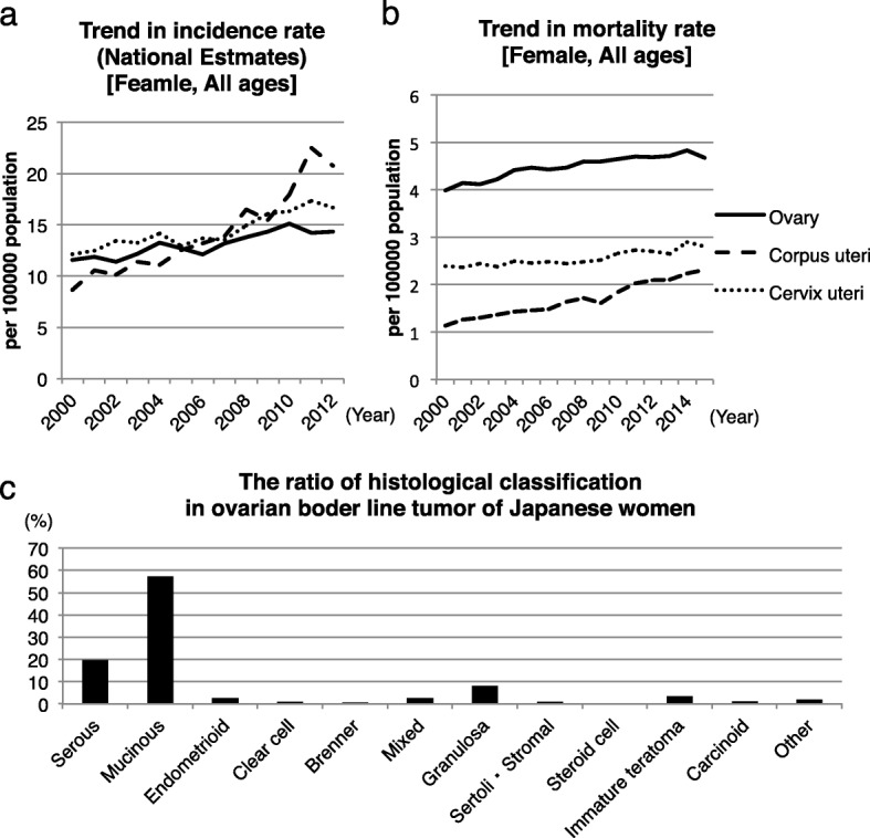
Background: The mortality rate of ovarian cancer is the highest among all gynecological malignancies in Japan. Ovarian tumors are classified as benign, borderline malignant, or malignant. Anticipating the histological subtype with imaging only is often difficult because of several histological subtypes of epithelial ovarian tumors (such as serous, mucinous, endometrioid, clear cell, and Brenner tumors). In addition, the majority of mucinous tumors in the ovary are metastatic. Furthermore, mucinous tumors belong to one of the two different subclasses (i.e., intestinal and seromucinous types). Ovarian seromucinous cystic tumors of borderline malignancy are infrequent and only rarely coexist with other malignant tumors.
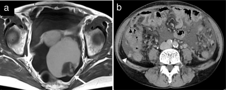
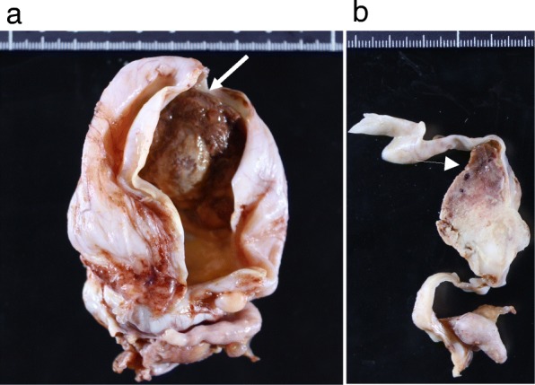
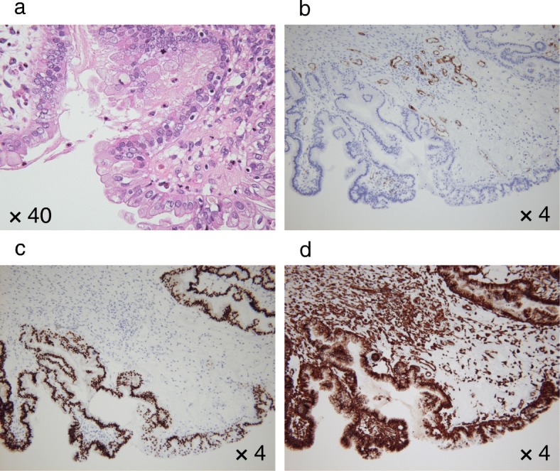
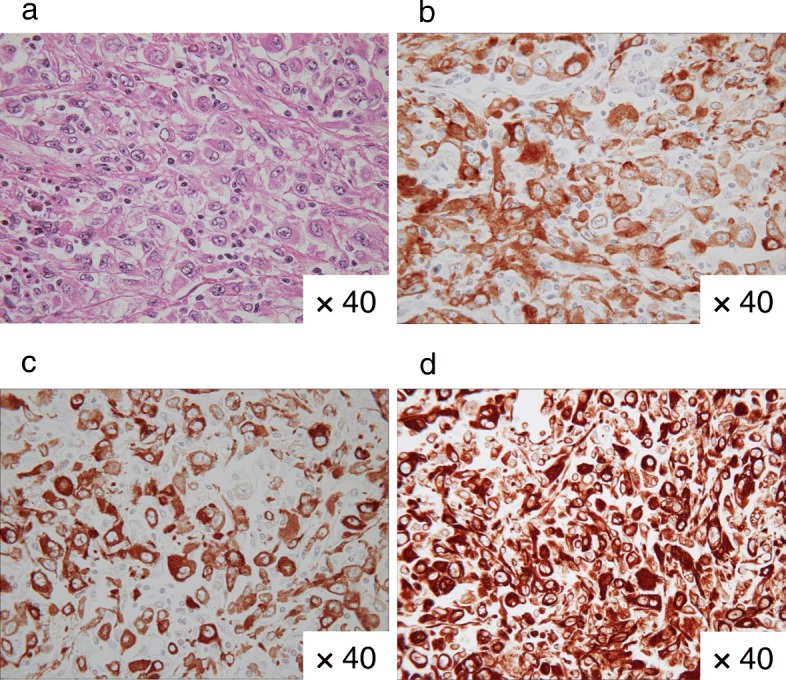
Case presentation: We have reported a 53-year-old Japanese woman with anaplastic carcinoma in an ovarian seromucinous cystic tumor of borderline malignancy. Her MRI and CT analysis revealed an ovarian tumor with a mural nodule, ascites, and peritoneal dissemination. Enhanced MRI revealed that the mural nodule was enhanced. Enhanced CT analysis revealed that the lymph nodes were not swollen. Intriguingly, the mural nodule crossed the cyst wall into the cavity and onto the surface. Her laboratory data revealed high serum CA 125 level. Cumulatively, these results suggested ovarian malignancy. The patient underwent hysterectomy with bilateral salpingo-oophorectomy, omentectomy, and resection of the disseminated lesions. Lymph node biopsy was omitted because of the suggestion of enhanced CT image findings and palpation during surgery. Her postoperative specimen examination determined FIGO at least stage IIIB, and accordingly, adjuvant chemotherapy was prescribed. After 3 years of the operation, the patient is presently alive without clinical tumor recurrences.
Conclusion: Imaging studies with pathognomonic findings contributed to ovarian cancer diagnosis in this case. To the best of our knowledge, this is the first study in English literature to report detailed classification of mucinous borderline malignancy, seromucinous cystic, and anaplastic carcinoma in an ovarian seromucinous cystic tumor of borderline malignancy.
Keywords: Anaplastic tumor; Case report; Immunohistochemical; Mural nodule; Ovarian tumor; Seromucinous borderline malignancy.
Conflict of interest statement
Ethics approval and consent to participate
This report was approved by the Ethics Committee of Maruyama Memorial General Hospital (No. 2017–2).
Consent for publication
Written informed consent was obtained from the patient for publication of this case report and any accompanying images.
Competing interests
The authors declare that they have no competing interests.
Publisher’s Note
Springer Nature remains neutral with regard to jurisdictional claims in published maps and institutional affiliations.
Figures
References
-
- Center for Cancer Control and Information Service, National Cancer Center, Japan. https://ganjoho.jp/reg_stat/statistics/dl/index.html.
-
- Buttin BM, Herzog TJ, Powell MA, Rader JS, Mutch DG. Epithelial ovarian tumors of low malignant potential: the role of microinvasion. Obstet Gynecol. 2002;99(1):11–17. - PubMed
-
- Kurman RJ, Carcangiu ML, Herrington CS, et al. WHO Classification of Tumours. 4. Aufl. Lyon: WHO Press; 2014. WHO Classification of Tumours of Female Reproductive Organs.
Publication types
MeSH terms
LinkOut - more resources
Full Text Sources
Other Literature Sources
Medical
Research Materials
