Review
. 2018 Sep;38(5):461-476.
doi: 10.1016/j.semnephrol.2018.05.016.

Aberrant Glycosylation of the IgA1 Molecule in IgA Nephropathy

Affiliations
Review

Aberrant Glycosylation of the IgA1 Molecule in IgA Nephropathy

Jan Novak et al. Semin Nephrol. 2018 Sep.

Abstract

IgA nephropathy, the most common primary glomerulonephritis in the world and a frequent cause of end-stage renal disease, is characterized by typical mesangial deposits of IgA1, as described by Berger and Hinglaise in 1968. Since then, it has been discovered that aberrant IgA1 O-glycosylation is involved in disease pathogenesis. Progress in glycomic, genomic, clinical, analytical, and biochemical studies has shown autoimmune features of IgA nephropathy. The autoimmune character of the disease is explained by a multihit pathogenesis model, wherein overproduction of aberrantly glycosylated IgA1, galactose-deficient in some O-glycans, by IgA1-secreting cells leads to increased levels of circulatory galactose-deficient IgA1. These glycoforms induce production of autoantibodies that subsequently bind hinge-region of galactose-deficient IgA1 molecules, resulting in the formation of nephritogenic immune complexes. Some of these complexes deposit in the kidney, activate mesangial cells, and incite glomerular injury. Thus, galactose-deficient IgA1 is central to the disease process. In this article, we review studies concerning IgA1 O-glycosylation that have contributed to the current understanding of the role of IgA1 in the pathogenesis of IgA nephropathy.

Keywords: IgA1; O-glycans; autoantibody; signaling.


Figures

Figure 1.
Figure 1.. Structure and O-glycosylation of human circulatory IgA1.
Modeled structure of glycosylated monomeric IgA1 based on PDB ID: 1IGA (top) and hinge-region amino acid sequence with attachment sites of six O-glycans (bottom). Modeled O-and N-glycans are depicted as spheres for clarity and are based on observed glycoforms: red for Gal-GalNAc; orange for GalNAc; and magenta for N-glycan (NeuAc)2(Gal)2(GlcNAc)2+(Fuc)1(Man)3(GlcNAc)2. Hinge-region non-glycosylated amino-acid residues are in blue. There are up to six sites with O-glycans in the IgA1 hinge region: at T225, T228, S230, S232, T233, and T236 (marked by stars in the bottom amino-acid sequence). Model was generated by Tyler Stewart; figure is reproduced with permission.
Figure 2.
Figure 2.. Site-specific microheterogeneity of normal human serum IgA1 hinge-region O-glycans.
This figure summarizes results based on relative distributions of desialylated O-glycopeptides observed by high-resolution mass spectrometry. This distribution includes the delineation of amino-acid positional isomers identified by liquid chromatography-mass spectrometry and ECD-tandem mass spectrometry. S230, T233 and T236 are the primary sites of Gal deficiency and variable positional isomers in normal serum IgA1. S225, S228, and S232 are predominantly occupied by Gal-GalNAc disaccharide; these sites are rarely non-glycosylated (N.G.) or occupied by Gal-deficient species. Figure is reproduced with permission.
Figure 3.
Figure 3.. Biosynthetic pathways of IgA1 O-glycosylation.
O-glycans are synthesized in a step-wise manner, beginning with the attachment of N-acetylgalactosamine (GalNAc) to the oxygen atom of serine (Ser) or threonine (Thr) that is catalyzed by GalNAc-transferases. This step can be followed by addition of galactose (Gal) (catalyzed by C1GalT1; chaperone Cosmc is required for production of active C1GalT1 enzyme). Sialic acid (N-acetylneuraminic acid) can be added to each glycan by different enzymes, ST3Gal-1 for sialylation of Gal and ST6GalNAc-II for sialylation of GalNAc. Sialylation of terminal GalNAc (i.e., without Gal) prevents subsequent addition of Gal. Pathway is proposed based on published primary data and reviews ,,,,,–. Color coding is per Symbol Nomenclature for Glycans: yellow-filled square, GalNAc; yellow-filled circle, Gal; purple-filled diamond, N-acetylneuraminic acid.
Figure 4.
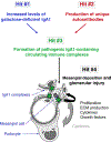
Figure 4.. A multi-hit hypothesis of IgAN pathogenesis.
This model assumes formation of immune complexes in the circulation and their subsequent mesangial deposition. An alternative hypothesis assumes that aberrantly glycosylated IgA1 deposits in the mesangium as lanthanic deposits that are later bound by newly appearing autoantibodies, resulting in the in situ formation of immune complexes. Hit 1: Increased production of Gal-deficient IgA1 by a sub-population of IgA1-secreting cells. Hit 2: Formation of autoantibodies with specific characteristics of the variable region of the heavy chain that recognize Gal-deficient IgA1. Hit 3: Formation of pathogenic immune complexes from auto-antigen (Gal-deficient IgA1) and Gal-deficient IgA1-specific autoantibody.,– Hit 4: Deposition of pathogenic immune complexes in the mesangium, activation of mesangial cells, and induction of glomerular injury.,,,,– These hits may be regulated by distinct genetic loci, identified by genome-wide association studies (GWAS) as associated with risk for IgAN as well as by other loci (for review see,). An alternative hypothesis assumes that aberrantly glycosylated IgA1 deposits in the mesangium as lanthanic deposits that are later bound by newly appearing autoantibodies, resulting in the in situ formation of immune complexes.

References

    1. Julian BA, Waldo FB, Rifai A, Mestecky J. IgA nephropathy, the most common glomerulonephritis worldwide. A neglected disease in the United States? Am J Med. 1988;84:129–32. - PubMed
    1. Berger J, Hinglais N. Les dépôts intercapillaires d’IgA-IgG (Intercapillary deposits of IgA-IgG). J Urol Nephrol. 1968;74:694–5. - PubMed
    1. Jennette JC. The immunohistology of IgA nephropathy. Am J Kidney Dis. 1988;12(5):348–52. - PubMed
    1. Conley ME, Cooper MD, Michael AF. Selective deposition of immunoglobulin A1 in immunoglobulin A nephropathy, anaphylactoid purpura nephritis, and systemic lupus erythematosus. J Clin Invest. 1980;66:1432–6. - PMC - PubMed
    1. Allen AC, Bailey EM, Brenchley PEC, Buck KS, Barratt J, Feehally J. Mesangial IgA1 in IgA nephropathy exhibits aberrant O-glycosylation: Observations in three patients. Kidney Int. 2001;60:969–73. - PubMed

Publication types

MeSH terms