Quantitative distribution of choline acetyltransferase activity in rat trapezoid body
- PMID: 30177425
- PMCID: PMC6240496
- DOI: 10.1016/j.heares.2018.08.008
Quantitative distribution of choline acetyltransferase activity in rat trapezoid body
Abstract
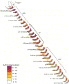
There is evidence for a function of acetylcholine in the cochlear nucleus, primarily in a feedback, modulatory effect on auditory processing. Using a microdissection and quantitative microassay approach, choline acetyltransferase activity was mapped in the trapezoid bodies of rats, in which the activity is relatively higher than in cats or hamsters. Maps of series of sections through the trapezoid body demonstrated generally higher choline acetyltransferase activity rostrally than caudally, particularly in its portion ventral to the medial part of the spinal trigeminal tract. In the lateral part of the trapezoid body, near the cochlear nucleus, activities tended to be higher in more superficial portions than in deeper portions. Calculation of choline acetyltransferase activity in the total trapezoid body cross-section of a rat with a comprehensive trapezoid body map gave a value 3-4 times that estimated for the centrifugal labyrinthine bundle, which is mostly composed of the olivocochlear bundle, in the same rat. Comparisons with other rats suggest that the ratio may not usually be this high, but it is still consistent with our previous results suggesting that the centrifugal cholinergic innervation of the rat cochlear nucleus reaching it via a trapezoid body route is much higher than that reaching it via branches from the olivocochlear bundle. The higher choline acetyltransferase activity rostrally than caudally in the trapezoid body is consistent with evidence that the centrifugal cholinergic innervation of the cochlear nucleus derives predominantly from locations at or rostral to its anterior part, in the superior olivary complex and pontomesencephalic tegmentum.
Keywords: Acetylcholine; Acetylcholinesterase; Auditory; Cochlear nucleus; Hearing.
Copyright © 2018 Elsevier B.V. All rights reserved.
Figures


Similar articles
-
Effect of olivocochlear bundle transection on choline acetyltransferase activity in the rat cochlear nucleus.Hear Res. 1987;28(2-3):237-51. doi: 10.1016/0378-5955(87)90052-9. Hear Res. 1987. PMID: 3654392
-
Contribution of centrifugal innervation to choline acetyltransferase activity in the cat cochlear nucleus.Hear Res. 1990 Nov;49(1-3):259-79. doi: 10.1016/0378-5955(90)90108-2. Hear Res. 1990. PMID: 2292500
-
Effects of cochlear ablation on choline acetyltransferase activity in the rat cochlear nucleus and superior olive.J Neurosci Res. 2005 Jul 1;81(1):91-101. doi: 10.1002/jnr.20536. J Neurosci Res. 2005. PMID: 15931674
-
Effects of trapezoid body and superior olive lesions on choline acetyltransferase activity in the rat cochlear nucleus.Hear Res. 1987;28(2-3):253-70. doi: 10.1016/0378-5955(87)90053-0. Hear Res. 1987. PMID: 3654393
-
Cholinergic cells of the pontomesencephalic tegmentum: connections with auditory structures from cochlear nucleus to cortex.Hear Res. 2011 Sep;279(1-2):85-95. doi: 10.1016/j.heares.2010.12.019. Epub 2010 Dec 30. Hear Res. 2011. PMID: 21195150 Free PMC article. Review.
Cited by
-
Amino acid and acetylcholine chemistry in mountain beaver cochlear nucleus and comparisons to pocket gopher, other rodents, and cat.Hear Res. 2020 Jan;385:107841. doi: 10.1016/j.heares.2019.107841. Epub 2019 Nov 10. Hear Res. 2020. PMID: 31765816 Free PMC article.
-
Chemical Effects of Kainic Acid Injection into the Rat Superior Olivary Region.HSOA J Otolaryngol Head Neck Surg. 2020;6(2):045. doi: 10.24966/ohns-010x/100045. Epub 2020 Jul 22. HSOA J Otolaryngol Head Neck Surg. 2020. PMID: 33733053 Free PMC article.
References
-
- Adams JC, 1989. Non-olivocochlear cholinergic periolivary cells. Soc. Neurosci. Abstr 15, abstr. 445.1.
-
- Brown JC, Howlett B, 1968. The facial outflow and the superior salivatory nucleus: an histochemical study in the rat. J. Comp. Neurol 134, 175–192. - PubMed
-
- Brown JC, Howlett B, 1971. Rat hindbrain thiocholine reactions relevant to the localization of the inferior salivatory nucleus. Acta Anat 79, 333–359. - PubMed
-
- Caspary DM, Havey DC, Faingold CL, 1983. Effects of acetylcholine on cochlear nucleus neurons. Exp. Neurol 82, 491–498. - PubMed
-
- Chen K, Waller HJ, Godfrey TG, Godfrey DA, 1999. Glutamatergic transmission of neuronal responses to carbachol in rat dorsal cochlear nucleus slices. Neuroscience 90, 1043–1049. - PubMed
Publication types
MeSH terms
Substances
Grants and funding
LinkOut - more resources
Full Text Sources
Other Literature Sources
Miscellaneous

