. 2018 Sep 3;9(1):3569.
doi: 10.1038/s41467-018-06038-y.

SCFFBW7-mediated degradation of Brg1 suppresses gastric cancer metastasis

Affiliations

SCFFBW7-mediated degradation of Brg1 suppresses gastric cancer metastasis

Li-Yu Huang et al. Nat Commun. .

Abstract

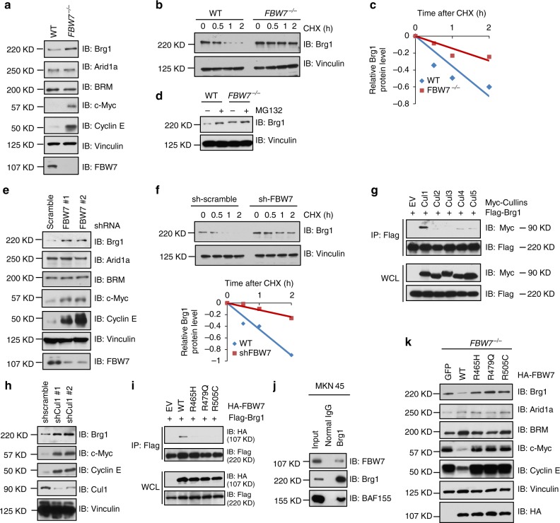
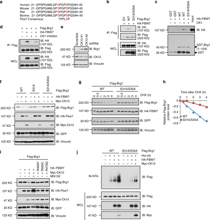
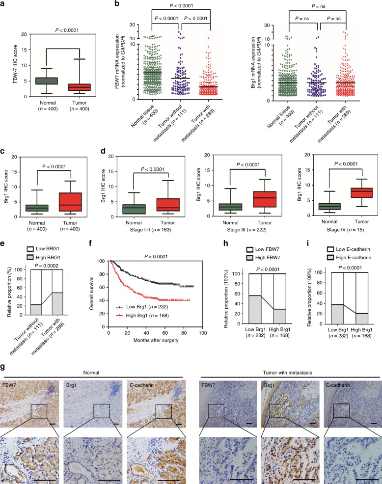
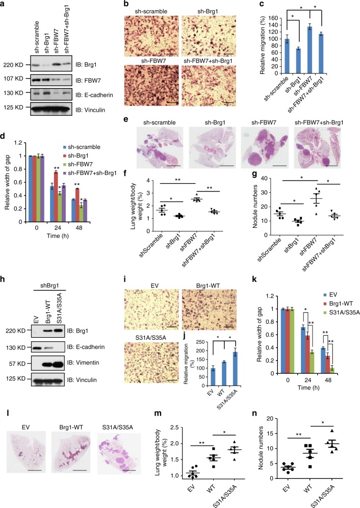
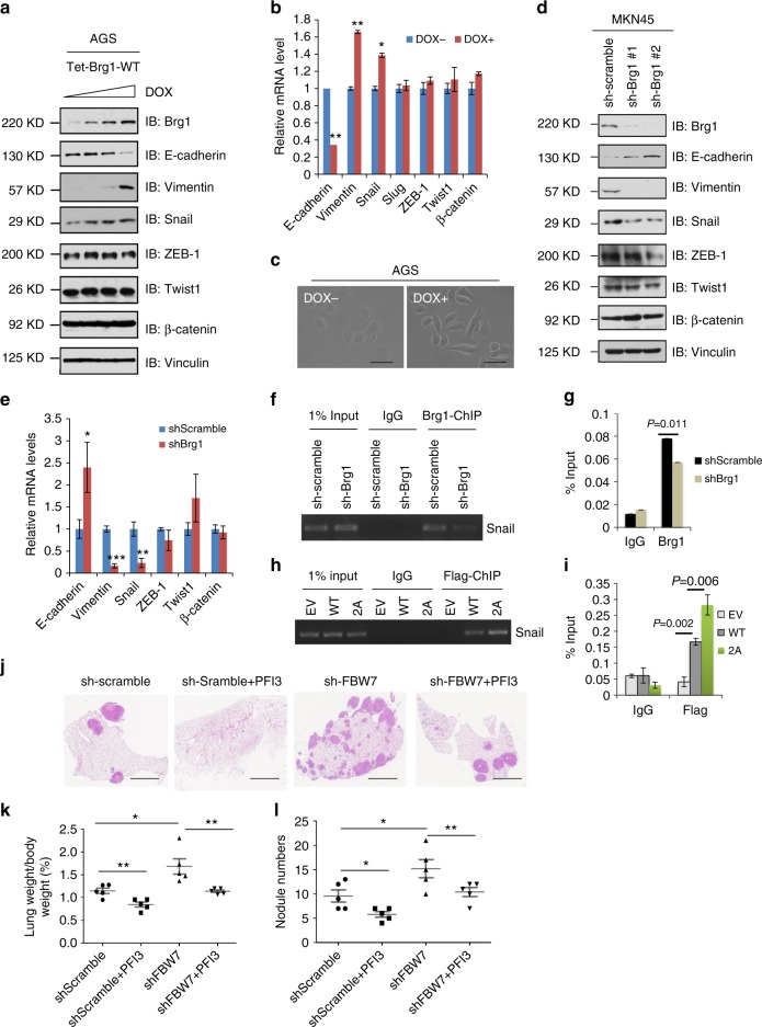
Brg1/SMARCA4 serves as the ATPase and the helicase catalytic subunit for the multi-component SWI/SNF chromatin remodeling complex, which plays a pivotal role in governing chromatin structure and gene transcription. However, the upstream signaling pathways regulating Brg1 protein stability and its physiological contribution to carcinogenesis remain largely elusive. Here we report that Brg1 is a bona fide ubiquitin substrate of SCFFBW7. We reveal that CK1δ phosphorylates Brg1 at Ser31/Ser35 residues to facilitate the binding of Brg1 to FBW7, leading to ubiquitination-mediated degradation. In keeping with a tumor suppressive role of FBW7 in human gastric cancer, we find an inverse correlation between FBW7 and Brg1 expression in human gastric cancer clinical samples. Mechanistically, we find that stabilization of Brg1 in gastric cancer cells suppresses E-cadherin expression, subsequently promoting gastric cancer metastasis. Hence, this previously unknown FBW7/Brg1 signaling axis provides the molecular basis and the rationale to target Brg1 in FBW7-compromised human gastric cancers.


Conflict of interest statement

The authors declare no competing interests.

Figures

Fig. 1
Fig. 1
FBW7 negatively regulates the stability of Brg1. a Immunoblot analysis (IB) of whole cell lysates (WCLs) derived from wild-type (WT) and FBW7/− DLD1 cells. b, c WT and FBW7−/− DLD1 cells were treated with 20 μg/ml cycloheximide (CHX). At the indicated time points, WCLs were prepared and IB analysis was carried out with indicated antibodies (b). The relative Brg1 intensity was normalized to Vinculin and then normalized to the t = 0 controls (c). d IB analysis of WCLs derived from WT and FBW7−/− DLD1 cells treated with MG132 (10 μM) or DMSO for 10 h. e IB analysis of WCLs derived from MKN45 cells infected with the indicated shRNA lentiviruses. f IB analysis of WCLs derived from MKN45 cells infected with the indicated shRNA lentiviruses and treated with 20 μg/ml CHX for indicated time periods. The relative intensity of Brg1 was normalized to Vinculin and then normalized to the t = 0 control (Bottom). g IB analysis of WCLs and immunoprecipitated (IPs) derived from 293T cells transfected with Flag-Brg1 together with the indicated constructs of Myc-tagged Cullin family members. h IB analysis of WCLs derived from MKN45 cells infected with the indicated lentiviral shRNA-Cullin1 constructs. i IB analysis of WCLs and IPs derived from 293T cells transfected with Flag-Brg1 together with the indicated FBW7 constructs. j Co-IP experiments in MKN45 cells were performed using anti-Brg1 antibody (sc-17796, Santa Cruz). Mouse IgG was used as a control. k IB analysis of WCLs derived from FBW7−/− DLD1 cells infected with the indicated FBW7-expressing lentiviral vectors
Fig. 2
Fig. 2
Brg1 interacts with FBW7 in a CKIδ-mediated phosphorylation dependent manner. a Sequence alignment of Brg1 with the phospho-degron sequences recognized by FBW7. The putative FBW7 phospho-degron sequence present in Brg1 is conserved across different species. b IB analysis of WCLs and IPs derived from 293T cells transfected with HA-FBW7 together with the indicated Flag-Brg1 constructs. c IB analysis showing the enhanced binding of HA-FBW7 with bacterially purified GST-Brg1 (1–104aa) proteins after incubation with CK1 kinase in vitro. GST was used as a negative control. d IB analysis of WCLs and IPs derived from 293T cells transfected with HA-FBW7 and Flag-Brg1. Where indicated, the CK1 inhibitor IC261 (20 μM) was added for 8 h before harvesting for IB analysis. DMSO was used as a negative control. e IB analysis of WCLs derived from MKN45 cells infected with the indicated lentiviral shRNA-CK1δ constructs. f IB analysis of WCLs derived from 293T cells transfected with the indicated Flag-Brg1 and HA-FBW7 vectors in the presence or absence of Myc-CK1δ. GFP was used as a negative control. g, h 293T cells were transfected with the indicated Flag-Brg1 constructs together with HA–FBW7 and Myc-CK1δ plasmids. Twenty hours after transfection, cells were split into 60-mm dishes. After another 20 h, cells were treated with 20 μg/ml CHX. i IB analysis of WCLs derived from 293T cells transfected with Flag-Brg1 and the indicated HA-FBW7 vectors. Where indicated, MG132 (10 μM) was treated for 10 h. j IB analysis of His tag pull-down and WCLs derived from 293 cells transfected with indicated Flag-Brg1 constructs together with the HA-FBW7, Myc-CK1δ, and His-Ub plasmids. 20 h after transfection, cells were treated with MG132 for 10 h before cell collection. His tag pull-down was then performed. Ni-NTA: nickel-nitrilotriacetic acid, Ub: ubiquitin
Fig. 3
Fig. 3
Brg1 expression inversely correlates with FBW7 to govern gastric cancer progression. a The scores of FBW7 immunohistochemical (IHC) staining in 400 tumor sections and paired adjacent non-tumor tissues. b The mRNA expressions of FBW7 and Brg1 were examined by real-time PCR. The data were analyzed by Student’s t test. c, d The scores of Brg1 immunohistochemical staining in tumor sections and paired adjacent non-tumor tissues in total (c) or in different TNM (tumor node metastasis) stages (d). The data were analyzed by Student’s t test. e High Brg1 expression positively correlated with lymph node metastasis in gastric cancer. Their correlation was analyzed by Spearman rank correlation test. f The association of Brg1 expression with overall survival was examined by the Kaplan–Meier analysis in gastric cancer patients from the Zhongshan cohort. g Representative images of FBW7, Brg1, and E-cadherin staining were shown in consecutive normal sections and distant metastatic tumor sections. Scale bar, 100 μm. h, i The correlations between Brg1 and FBW7 levels (h), Brg1 and E-cadherin levels (i) in 400 consecutive tumor sections that were semiquantified as high or low and analyzed by Spearman rank correlation test
Fig. 4
Fig. 4
Brg1 promotes cell migration in vitro and metastasis in vivo in gastric cancer cells. a IB analysis of WCLs derived from MKN45 cells infected with the indicated lentiviral shRNA constructs. b, c Representative images of migrated MKN45 cells infected with the indicated lentiviral shRNA constructs in a transwell assay (b) and quantification of migrated cells in (c). Data were shown as mean ± SD of three independent experiments. *p < 0.05, Student’s t test. Scale bar, 100 μm. d The migration of MKN45 cells transfected with the indicated lentiviral shRNA plasmids in scratch experiments. Data were shown as mean ± SD of three independent experiments. *p < 0.05, **p < 0.01, Student’s t test. eg Representative images and statistical analysis of metastasis formed in lungs of each nude mouse (n = 5) at 2 months after tail vein injection of MKN45 cells (3 × 106) with the indicated lentiviral shRNA plasmids. Scale bar, 1 mm. h IB analysis of whole cell lysates derived from AGS cells transfected with the indicated Flag-Brg1 constructs. i, j Representative images of migrated AGS cells transfected with the indicated Flag-Brg1 constructs in a transwell assay and quantification of migrated cells. Data were shown as mean ± SD of three independent experiments. *p < 0.05, Student’s t test. Scale bar, 100 μm. k The migration of AGS cells transfected with the indicated Flag-Brg1 constructs in scratch experiments. Data are shown as mean ± SD of three independent experiments. *p < 0.05, **p < 0.01, Student’s t test. ln Representative images and statistical analysis of metastases that formed in lungs of each nude mouse (n = 5) at 2 months after tail vein injection of AGS cells (2 × 106) transfected with the indicated Flag-Brg1 plasmids. *p < 0.05, **p < 0.01, Student’s t test. Scale bar, 1 mm
Fig. 5
Fig. 5
Brg1 promotes metastasis in gastric cancer by driving an EMT transcriptional program. a IB analysis of WCLs derived from gastric cancer cell lines AGS. AGS cells were infected with the indicated pTRIPZ-Brg1 lentiviral vectors and were selected with 1 μg/ml puromycin for 72 h to eliminate non-infected cells. Afterward, 100, 300, 1000, 3000 ng/ml doxycycline (DOX) was added to the cells for another 24 h before harvesting for IB analysis. b Real-time PCR assays were performed in pTRIPZ-Brg1 AGS cells in the present or absence of DOX (1000 ng/ml) for 24 h. *p < 0.05, **p < 0.01, Student’s t test. c Representative images of the cell morphology in AGS cells. Scale bar, 100 μm. d IB analysis of WCLs derived from MKN45 cells infected with the indicated lentiviral shRNA plasmids. e Real-time PCR assays were performed in MKN45 cells infected with the indicated lentiviral shRNA plasmids.*p < 0.05, **p < 0.01, Student’s t test. f, g ChIP assays with anti-Brg1 antibody were performed in MKN45 cells infected with the indicated lentiviral shRNA plasmids. Mouse IgG was used as negative control. h, i ChIP assays with anti-Flag antibody were performed in AGS cells transfected with the indicated Flag-Brg1 constructs. Mouse IgG was used as antibody control in this experiment. jl Representative images and statistical analysis of metastases that formed in lungs of each nude mouse (n = 5) at 2 months after tail vein injection of MKN45 cells (3 × 106/each) transfected with the indicated lentiviral shRNA plasmids. One milligrams PFI3 (SIGMA, SML0939) dissolved in dimethyl sulfoxide plus 0.25 ml phosphate-buffered saline was given intraperitoneally 3 times per week. *p < 0.05, **p < 0.01, Student’s t test. Scale bar, 1 mm
Fig. 6
Fig. 6
The proposed model for FBW7 in suppressing metastasis in gastric cancer setting. The FBW7/Brg1 signaling axis governs gastric cancer EMT changes and plays a critical role in gastric cancer metastasis

References

    1. Peterson CL, Tamkun JW. The SWI-SNF complex: a chromatin remodeling machine? Trends Biochem. Sci. 1995;20:143–146. doi: 10.1016/S0968-0004(00)88990-2. - DOI - PubMed
    1. Whitehouse I, et al. Nucleosome mobilization catalysed by the yeast SWI/SNF complex. Nature. 1999;400:784–787. doi: 10.1038/23506. - DOI - PubMed
    1. Kassabov SR, Zhang B, Persinger J, Bartholomew B. SWI/SNF unwraps, slides, and rewraps the nucleosome. Mol. Cell. 2003;11:391–403. doi: 10.1016/S1097-2765(03)00039-X. - DOI - PubMed
    1. Wilson BG, Roberts CW. SWI/SNF nucleosome remodellers and cancer. Nat. Rev. Cancer. 2011;11:481–492. doi: 10.1038/nrc3068. - DOI - PubMed
    1. Reisman DN, Sciarrotta J, Wang W, Funkhouser WK, Weissman BE. Loss of BRG1/BRM in human lung cancer cell lines and primary lung cancers: correlation with poor prognosis. Cancer Res. 2003;63:560–566. - PubMed

Publication types