

Gulf war illness-related chemicals increase CD11b/c+ monocyte infiltration into the liver and aggravate hepatic cholestasis in a rodent model
- PMID: 30177688
- PMCID: PMC6120951
- DOI: 10.1038/s41598-018-31599-9
Gulf war illness-related chemicals increase CD11b/c+ monocyte infiltration into the liver and aggravate hepatic cholestasis in a rodent model
Abstract
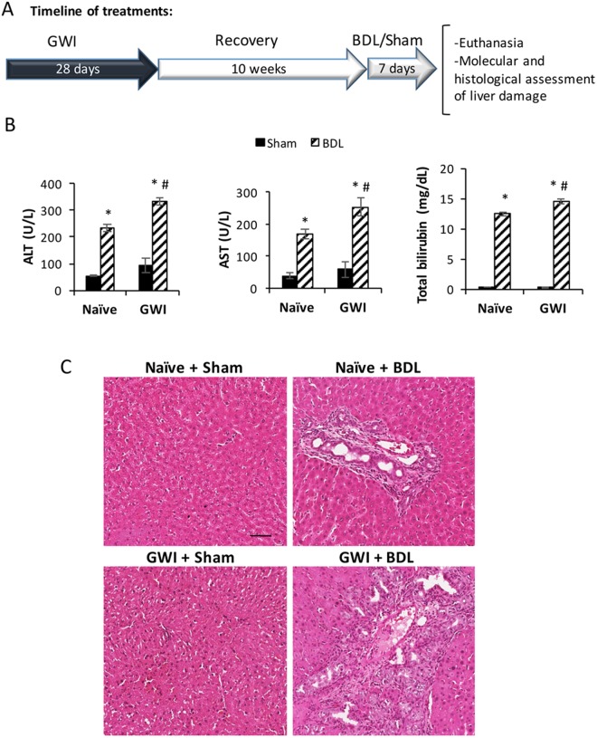
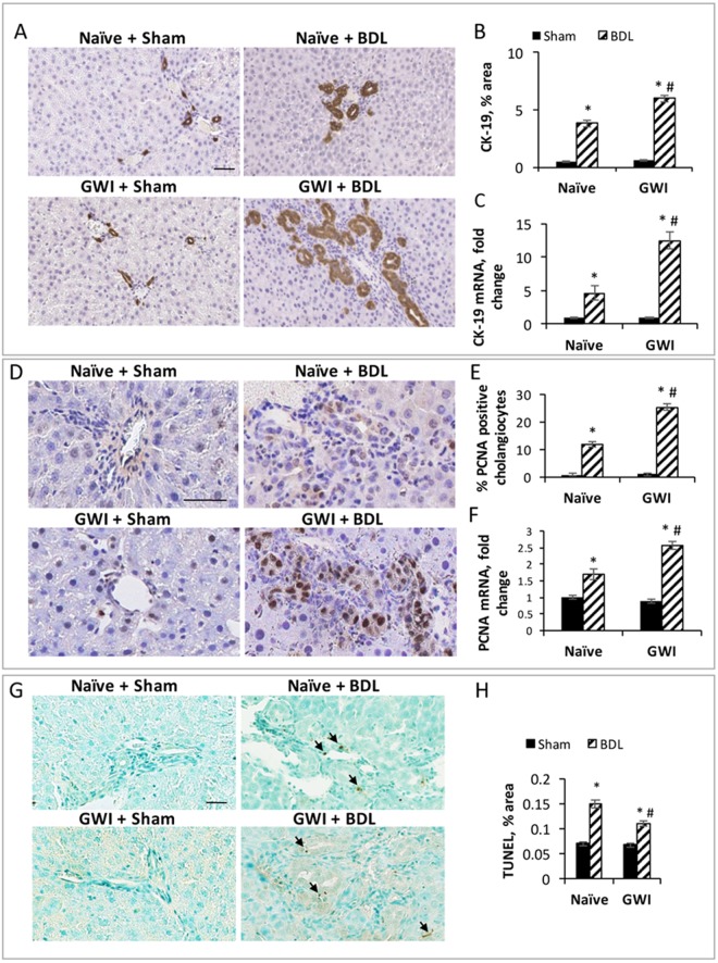
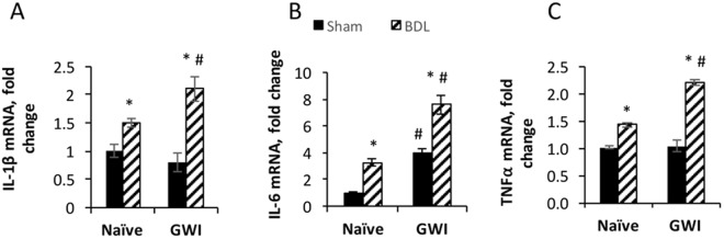
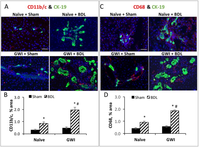
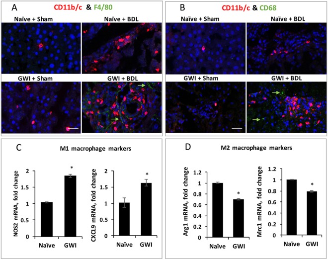
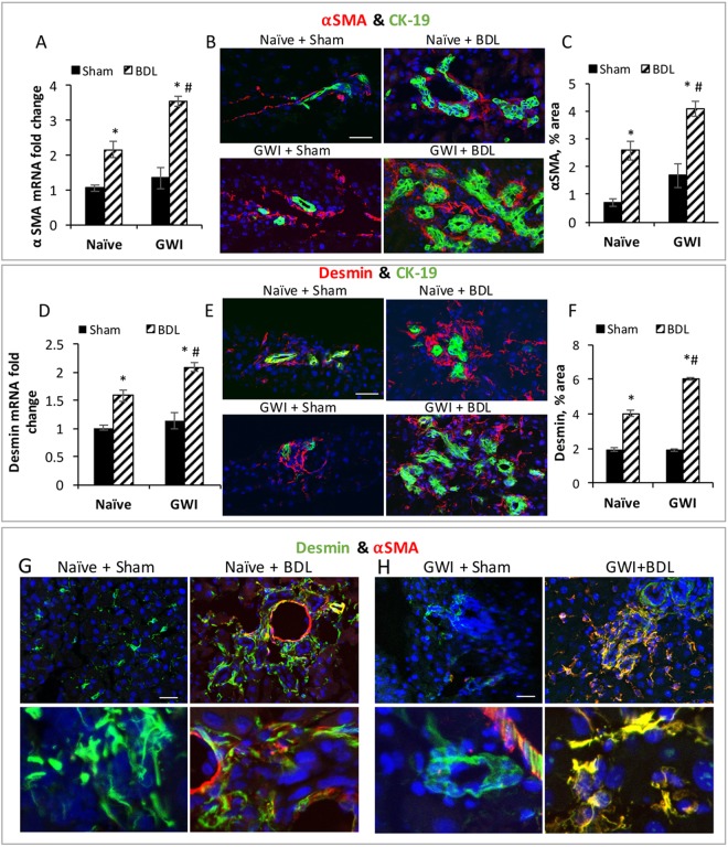
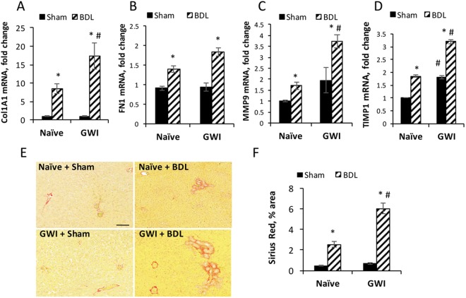
Gulf War Illness (GWI) is a chronic multisymptom disorder affecting veterans of the 1990-91 Gulf war. GWI was linked with exposure to chemicals including the nerve gas prophylactic drug pyridostigmine-bromide (PB) and pesticides (DEET, permethrin). Veterans with GWI exhibit prolonged, low-level systemic inflammation, though whether this impacts the liver is unknown. While no evidence exists that GWI-related chemicals are hepatotoxic, the prolonged inflammation may alter the liver's response to insults such as cholestatic injury. We assessed the effects of GWI-related chemicals on macrophage infiltration and its subsequent influence on hepatic cholestasis. Sprague Dawley rats were treated daily with PB, DEET and permethrin followed by 15 minutes of restraint stress for 28 days. Ten weeks afterward, GWI rats or naïve age-matched controls underwent bile duct ligation (BDL) or sham surgeries. Exposure to GWI-related chemicals alone increased IL-6, and CD11b+F4/80- macrophages in the liver, with no effect on biliary mass or hepatic fibrosis. However, pre-exposure to GWI-related chemicals enhanced biliary hyperplasia and fibrogenesis caused by BDL, compared to naïve rats undergoing the same surgery. These data suggest that GWI patients could be predisposed to developing worse liver pathology due to sustained low-level inflammation of the liver when compared to patients without GWI.
Conflict of interest statement
The authors declare no competing interests.
Figures
Similar articles
-
Exposure to Gulf war illness-related chemicals exacerbates alcohol-induced liver damage in rodents.Sci Rep. 2024 Jul 1;14(1):14981. doi: 10.1038/s41598-024-65638-5. Sci Rep. 2024. PMID: 38951546 Free PMC article.
-
Mood and memory deficits in a model of Gulf War illness are linked with reduced neurogenesis, partial neuron loss, and mild inflammation in the hippocampus.Neuropsychopharmacology. 2013 Nov;38(12):2348-62. doi: 10.1038/npp.2013.158. Epub 2013 Jun 28. Neuropsychopharmacology. 2013. PMID: 23807240 Free PMC article.
-
Neurochemical and neuroinflammatory perturbations in two Gulf War Illness models: Modulation by the immunotherapeutic LNFPIII.Neurotoxicology. 2020 Mar;77:40-50. doi: 10.1016/j.neuro.2019.12.012. Epub 2019 Dec 19. Neurotoxicology. 2020. PMID: 31866310 Free PMC article.
-
Dysbiosis in gastrointestinal pathophysiology: Role of the gut microbiome in Gulf War Illness.J Cell Mol Med. 2023 Apr;27(7):891-905. doi: 10.1111/jcmm.17631. Epub 2023 Jan 30. J Cell Mol Med. 2023. PMID: 36716094 Free PMC article. Review.
-
Progression of intervention-focused research for Gulf War illness.Mil Med Res. 2019 Oct 18;6(1):31. doi: 10.1186/s40779-019-0221-x. Mil Med Res. 2019. PMID: 31627737 Free PMC article. Review.
Cited by
-
The mediating roles of obesity indicators and serum albumin in the association of DEET exposure with depression and sleep disorders in adults: evidence from NHANES 2007-2016.BMC Public Health. 2025 May 6;25(1):1658. doi: 10.1186/s12889-025-22880-4. BMC Public Health. 2025. PMID: 40329240 Free PMC article.
-
Industrial, Biocide, and Cosmetic Chemical Inducers of Cholestasis.Chem Res Toxicol. 2019 Jul 15;32(7):1327-1334. doi: 10.1021/acs.chemrestox.9b00148. Epub 2019 Jun 18. Chem Res Toxicol. 2019. PMID: 31243985 Free PMC article.
-
Gulf War Illness: Mechanisms Underlying Brain Dysfunction and Promising Therapeutic Strategies.Pharmacol Ther. 2021 Apr;220:107716. doi: 10.1016/j.pharmthera.2020.107716. Epub 2020 Oct 24. Pharmacol Ther. 2021. PMID: 33164782 Free PMC article. Review.
-
Exposure to Gulf war illness-related chemicals exacerbates alcohol-induced liver damage in rodents.Sci Rep. 2024 Jul 1;14(1):14981. doi: 10.1038/s41598-024-65638-5. Sci Rep. 2024. PMID: 38951546 Free PMC article.
-
Exposure to Gulf war illness-related chemicals exacerbates alcohol- induced liver damage in rodents.Res Sq [Preprint]. 2024 Jan 18:rs.3.rs-3838282. doi: 10.21203/rs.3.rs-3838282/v1. Res Sq. 2024. Update in: Sci Rep. 2024 Jul 1;14(1):14981. doi: 10.1038/s41598-024-65638-5. PMID: 38313276 Free PMC article. Updated. Preprint.
References
-
- United States. Department of Veterans Affairs. Research Advisory Committee on Gulf War Veterans’ Illnesses. Scientific progress in understanding Gulf War veterans’ illnesses: report and recommendations, (Dept. of Veterans Affairs, Research Advisory Committee on Gulf War Veterans’ Illnesses, Washington, D.C., 2004).
Publication types
MeSH terms
Substances
Grants and funding
LinkOut - more resources
Full Text Sources
Other Literature Sources
Medical
Research Materials
