. 2018 Sep 3;8(1):13140.
doi: 10.1038/s41598-018-31441-2.

Plasma gelsolin promotes re-epithelialization

Affiliations

Plasma gelsolin promotes re-epithelialization

J Wittmann et al. Sci Rep. .

Abstract

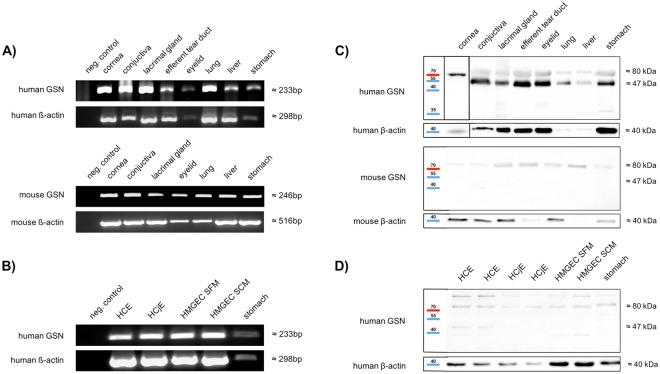
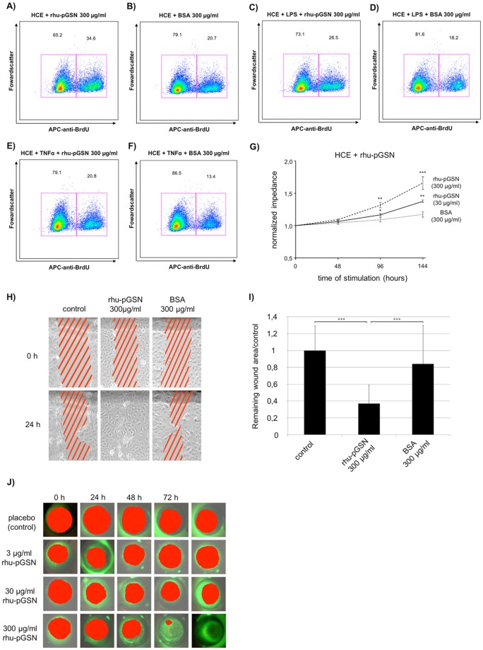
Woundhealing disorders characterized by impaired or delayed re-epithelialization are a serious medical problem that is painful and difficult to treat. Gelsolin (GSN), a known actin modulator, supports epithelial cell regeneration and apoptosis. The aim of this study was to estimate the potential of recombinant gelsolin (rhu-pGSN) for ocular surface regeneration to establish a novel therapy for delayed or complicated wound healing. We analyzed the influence of gelsolin on cell proliferation and wound healing in vitro, in vivo/ex vivo and by gene knockdown. Gelsolin is expressed in all tested tissues of the ocular system as shown by molecular analysis. The concentration of GSN is significantly increased in tear fluid samples of patients with dry eye disease. rhu-pGSN induces cell proliferation and faster wound healing in vitro as well as in vivo/ex vivo. TGF-β dependent transcription of SMA is significantly decreased after GSN gene knockdown. Gelsolin is an inherent protein of the ocular system and is secreted into the tear fluid. Our results show a positive effect on corneal cell proliferation and wound healing. Furthermore, GSN regulates the synthesis of SMA in myofibroblasts, which establishes GSN as a key protein of TGF-β dependent cell differentiation.


Conflict of interest statement

The authors declare no competing interests.

Figures

Figure 1
Figure 1
RT-PCR and Western blot analysis of GSN expression. (A,B) Expression of specific GSN mRNA amplification products in human (cornea (n = 3), conjunctiva (n = 3), lacrimal gland (n = 3), efferent tear duct (n = 3), eyelid (n = 3), lung (n = 3), liver (n = 1) and stomach (n = 1)) and mouse tissues (cornea (n = 6), conjunctiva (n = 6), lacrimal gland (n = 6), eyelid (n = 6), lung (n = 5), liver (n = 6) and stomach (n = 5)) and three different human cell lines (i.e. HCE = human cornea epithelial cells (n = 3); HCjE = human conjunctiva epithelial cells (n = 3); HMGEC SFM = human meibomian gland epithelial cells cultured with serum-free medium (n = 3); HMGEC SCM = human meibomian gland epithelial cells cultured with serum containing medium to induce differentiation (n = 3). All negative controls without template cDNA. Stomach, liver and lung served as positive controls. (C,D) Western blot analysis of human (cornea (n = 3), conjunctiva (n = 3), lacrimal gland (n = 3), efferent tear duct (n = 3), eyelid (n = 3), lung (n = 3), liver (n = 1) and stomach (n = 1)) and mouse tissue (cornea (n = 6), conjunctiva (n = 6), lacrimal gland (n = 6), eyelid (n = 6), lung (n = 5), liver (n = 6) and stomach (n = 5)) samples and three different human cell lines (HCE = human cornea epithelial cells (n = 3); HCjE = human conjunctiva epithelial cells (n = 3); HMGEC SFM = human meibomian gland epithelial cells cultured with serum free medium (n = 3); HMGEC SCM = human meibomian gland epithelial cells cultured with serum containing medium to induce differentiation (n = 3)) using an anti-GSN antibody. Stomach, liver and lung tissue samples served as positive controls.
Figure 2
Figure 2
Localization and quantification of GSN in tissues of the ocular surface and lacrimal apparatus as well as in healthy tears and tears from patients suffering from aqueous-deficient and evaporative forms of dry eye disease. Immunohistochemical localization of GSN in human and murine cornea (1A,2B), conjunctiva (2A,2B) meibomian gland (3A,3B) lacrimal gland (4A,4B), stomach (5A,5B) and lung (6A,6B). Stomach and lung are used as positive controls. Red staining indicates positive reactivity of the antibody. The right part of each picture shows magnification. Scale bar: [1A],[2A],[1B],[2B],[4B],[6B] 20 µm, [3A]–[6A] 50 µm, [3B],[5B] 100 µm. (C) Immunofluorescence detection of GSN in a human meibomian gland and a human lacrimal gland. (GSN: green; DAPI (4′,6-diamidino-2-phenylindole): blue). Scale bar: meibomian gland 200 µm, lacrimal gland 100 µm. (D) Western blot analysis of human tear fluid of healthy male and a female donors (each n = 1) using an anti-GSN antibody. (E,F) Quantification of GSN protein by enzyme-linked immunosorbent assay (ELISA) of human (E) and mouse (F) tissue samples. (E) Quantification of GSN in human tissues ELISA. Amount of GSN is in relation to total protein amount. (F) Quantification of GSN in mouse tissue and tear fluid by ELISA. (G) Quantification of GSN in tear fluid by ELISA. Analyzed samples were from healthy (n = 10), aqueous-deficient dry eye (ADDE) (n = 14) and hyperevaporation dry eye (EDE) (n = 14). Statistical significance: **p ≤ 0.005, ***p ≤ 0.0005.
Figure 3
Figure 3
Proliferation and wound healing studies. (AF) Cell proliferation of human corneal epithelial cells (HCE cell line) after treatment with recombinant human plasma gelsolin (rhu-pGSN) assessed with fluorescence-activated cell sorting (FACS). (A) Stimulation with rhu-pGSN and (B) devoid of rhu-pGSN (bovine serum albumin (BSA) protein control). (C,E) Stimulation with lipopolysaccharide (LPS) and tumor necrosis factor α (TNFα) in combination with rhu-pGSN. (D,F) Stimulation with LPS and TNFα in combination with BSA. (G) Normalized impedance after stimulation with rhu-pGSN assessed with Electric Cell-Substrate Impedance Sensing (ECIS®). Stimulations were BSA, 30 µg/ml rhu-pGSN, 300 µg/ml rhu-pGSN. Start of stimulation 0 hours. Statistical significance: *p≤0.05, **p≤0.005, ***p≤0.0005. (H) Scratch assay (n = 3) on HCE. Cells were wounded using a pippet tip. Wounded areas (red stripes) after 0 hours and 24 hours of incubation. (I) Restored wound area after scratch (n = 3) and 24 hour incubation with rhu-pGSN or BSA, compared to control values. The wound healing rates were significantly higher under stimulation with 300 µl/ml rhu-pGSN compared with no rhu-pGSN control as well as BSA protein control. Statistical significance: ***p < 0.001. (J) rhu-pGSN promotes re-epithelialization of corneal wounds in combined in vivo/ex vivo model. The measured wound areas are highlighted in red.
Figure 4
Figure 4
GSN dependent differentiation. (A) Representative pictures of cultivated human corneal fibroblasts without stimulation (=control) and after stimulation with rhuTGF-β (3 ng/ml), rhu-pGSN (300 µg/ml) or rhuTGF-β+rhu-pGSN (3 ng/ml + 300 µg/ml) in combination. (B) Immunofluorescence of SMA in non-stimulated (=control) and stimulated (either 3 ng/ml rhuTGF-β or 300 µg/ml rhu-pGSN or both in combination) human primary corneal fibroblasts. Unstimulated fibroblasts (=control) only reveal weak SMA reactivity. (C) Western blot analysis of unstimulated (=control) and stimulated corneal fibroblasts after stimulation with rhuTGF-β (3 ng/ml), rhu-pGSN (300 µg/ml), or rhuTGF-β + rhu-pGSN (3 ng/ml + 300 µg/ml) using anti-SMA antibody. All samples show a clear band at approx. 40 kDa. GAPDH served as loading control. (D) Semi-quantification of relative SMA synthesis from Fig. 4C. Columns are normalized to not stimulated fibroblasts (=control). (E) Results of realtime-PCR analysis of SMA-mRNA expression after gene knockdown with siRNA for GSN. The columns are normalized to measured value of group without GSN-siRNA and treated with 3 ng/ml rhuTGF-β. (F) Results of semi-quantification of relative SMA synthesis in Western blot analysis (data not shown) after GSN-siRNA gene knockdown. Column is normalized to measured values of human primary fibroblasts treated only with rhuTGF-β. Statistical significance: ns = not significant. Statistical significance: **p < 0.01,***p < 0.005, ****p < 0.0001.

References

    1. Drug-induced corneal damage. Prescrire Int23, 97–100 (2014). - PubMed
    1. Guo S, Dipietro LA. Factors affecting wound healing. J Dent Res. 2010;89:219–229. doi: 10.1177/0022034509359125. - DOI - PMC - PubMed
    1. Tuan TL, Nichter LS. The molecular basis of keloid and hypertrophic scar formation. Molecular medicine today. 1998;4:19–24. doi: 10.1016/S1357-4310(97)80541-2. - DOI - PubMed
    1. Pascolini D, Mariotti SP. Global estimates of visual impairment: 2010. The British journal of ophthalmology. 2012;96:614–618. doi: 10.1136/bjophthalmol-2011-300539. - DOI - PubMed
    1. Aghadoost D. Ocular trauma: an overview. Archives of trauma research. 2014;3:e21639. doi: 10.5812/atr.21639. - DOI - PMC - PubMed

Publication types

MeSH terms