. 2018 Oct;3(10):1175-1185.
doi: 10.1038/s41564-018-0230-7. Epub 2018 Sep 3.

Global spread of three multidrug-resistant lineages of Staphylococcus epidermidis

Affiliations

Global spread of three multidrug-resistant lineages of Staphylococcus epidermidis

Jean Y H Lee et al. Nat Microbiol. 2018 Oct.

Abstract

Staphylococcus epidermidis is a conspicuous member of the human microbiome, widely present on healthy skin. Here we show that S. epidermidis has also evolved to become a formidable nosocomial pathogen. Using genomics, we reveal that three multidrug-resistant, hospital-adapted lineages of S. epidermidis (two ST2 and one ST23) have emerged in recent decades and spread globally. These lineages are resistant to rifampicin through acquisition of specific rpoB mutations that have become fixed in the populations. Analysis of isolates from 96 institutions in 24 countries identified dual D471E and I527M RpoB substitutions to be the most common cause of rifampicin resistance in S. epidermidis, accounting for 86.6% of mutations. Furthermore, we reveal that the D471E and I527M combination occurs almost exclusively in isolates from the ST2 and ST23 lineages. By breaching lineage-specific DNA methylation restriction modification barriers and then performing site-specific mutagenesis, we show that these rpoB mutations not only confer rifampicin resistance, but also reduce susceptibility to the last-line glycopeptide antibiotics, vancomycin and teicoplanin. Our study has uncovered the previously unrecognized international spread of a near pan-drug-resistant opportunistic pathogen, identifiable by a rifampicin-resistant phenotype. It is possible that hospital practices, such as antibiotic monotherapy utilizing rifampicin-impregnated medical devices, have driven the evolution of this organism, once trivialized as a contaminant, towards potentially incurable infections.


Conflict of interest statement

Competing interests

The authors declare no competing interests.

Figures
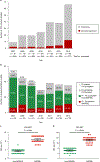
Fig. 1
Fig. 1. Increasing prevalence of multidrug-resistant S. epidermidis (MDRSe).
The prevalence of antibiotic-resistant S. epidermidis isolates at a single Australian institution over a seven-year period is shown. Susceptibilities were performed on a Vitek 2 (bioMérieux) platform and interpreted using CLSI criteria. a, Prevalence of teicoplanin (TEC)-resistant isolates. b, Prevalence of clinically significant isolates exhibiting rifampicin (R) and fusidic acid (FA) resistance. c,d, Vancomycin (VAN) (c) and teicoplanin (TEC) (d) METs were performed on 70 clinically significant S. epidermidis strains (35 rifampicin- and fusidic acid-susceptible (non-MDRSE); 35 rifampicin-plus fusidic acid-resistant (MDRSE)) plus two non-MDRSE reference isolates (RP62a and ATCC 12228); n = 72. The y axis is plotted on a log2 scale. Error bars represent geometric mean + 95% confidence interval (CI). Null hypothesis (no difference between means) was rejected for P < 0.05 (two-tailed Mann–Whitney U test of log2 transformed MICs).
Fig. 2
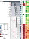
Fig. 2. Clonal expansion of endemic, multidrug-resistant ST2 and ST23 S. epidermidis lineages resulting in clinical disease within a single institution.
Maximum-likelihood core-SNP-based phylogeny for 69 clinically significant strains collected from 2007 to 2013 (34 multidrug S. epidermidis (MDRSE); 35 non-MDRSE); four closed, published S. epidermidis genomes; and the closed BPH0662 genome from the MDRSE index case (used as the reference strain); n = 74. Overlaid are the results of in silico MLST, BAPS, the determinants of rifampicin (RpoB mutations) and fusidic acid (fusB) resistance, and heatmap of vancomycin heteroresistant phenotype testing, as determined by VAN and TEC METs. The scale bar indicates number of nucleotide substitutions per site (bold), with an approximation of SNP rate (in parentheses). *Published reference strain SEI was not classifiable by the existing MLST scheme.
Fig. 3
Fig. 3. International clonal expansion of endemic, multidrug-resistant ST2 and ST23 S. epidermidis lineages resulting in clinical disease.
a, Maximum-likelihood, core-SNP-based phylogeny of 227 clinical isolates originating from 77 institutions in 10 countries, using BPH0662 as the reference genome, with BAPS groups overlaid. b, Subset analyses of the 133 ST2 isolates (plus ST188, a single locus variant of ST2). Insets show subset analyses for each of the four main MDRSE lineages: ST2-mixed (n = 60); ST2 BPH0662 clones (n = 71); ST23 (n = 50); ST5 (n = 15). Overlaid are the results of the determinants of rifampicin (RpoB mutations) and fusidic acid (fusB) resistance, and heatmap of vancomycin heteroresistant phenotype testing, as determined by VAN and TEC METs. BPH0662 was used as reference for all core-SNP subset analyses. Scale bars indicate number of nucleotide substitutions per site (bold), with an approximation of SNP rate (in parentheses). c, Pairwise-SNP analyses of the four main MDRSE lineages (sample sizes as in b).
Fig. 4
Fig. 4. RpoB mutations confer vancomycin heteroresistance in S. epidermidis.
a, An example of a VGA for BPH0676-WT, BPH0676-rpoB662 and the BPH0662 control strain. b, Biological triplicate data for the BPH0676-WT and BPH0676-rpoB662 mutant pair compared by VGA. Error bars represent mean ± 95% CI of three independent experiments. Null hypothesis (no difference between means) was rejected for P < 0.05 (two-tailed, unpaired Student’s t-test). c, Data for a single set of VAN and TEC METs for BPH0676-WT and the BPH0676-rpoB662 mutant pair. The y axis for METs is plotted on a log2 scale. d, Biological triplicate data for the BPH0676-WT and BPH0676-rpoB662 mutant pair compared by vancomycin PAP. Error bars represent mean ± 95% CI of three independent experiments. Null hypothesis (no difference between means) was rejected for P < 0.05 (two-tailed, unpaired Student’s t-test).
Fig. 5
Fig. 5. RpoB D471e and I527M mutations cause vancomycin heteroresistance in four different S. epidermidis backgrounds.
a–d, RpoB D471E and I527M dual mutations were introduced into four different, rifampicin-susceptible S. epidermidis strains: two clinically significant ST2 strains, BPH0676 and BPH0736, and reference strains RP62a (ST10) and ATCC 12228 (ST8). The gains in vancomycin tolerance of these rpoB662 mutants compared to their respective WT parental strains were individually validated (Supplementary Fig. 4), then summary data analysed. Three different phenotypic methods for the detection of vancomycin heteroresistance were used: VGA (a), VAN PAP (b), and VAN (c) and TEC (d) METs. The y axis for METs is plotted on a log2 scale. Data points for VGA and VAN PAP represent the mean of three independent experiments for each strain. Null hypothesis (no difference between means) was rejected for P < 0.05 (two-tailed, paired Student’s t-test).
Fig. 6
Fig. 6. Mutants containing dual D471e and I527M RpoB substitutions outcompete WT S. epidermidis in the presence of vancomycin.
Competition assays for the BPH0736-WT and rpoB662 mutant pair performed in the presence of VAN 0, 2 and 4 μg ml−1, with the percentage of rifampicin resistant (RIFR) to rifampicin susceptible (RIFS) isolates determined by plate count. All data points for biological triplicate experiments are displayed. Horizontal lines depict mean and error bars show standard deviation. Null hypothesis (no difference between means) was rejected for P < 0.05. Differences assessed using two-way analysis of variance (ANOVA) with Tukey’s test for multiple comparisons. ***P < 0.001, ****P < 0.0001.

Comment in

  • Time for biocide stewardship?
    Zamudio R, Oggioni MR, Gould IM, Hijazi K. Zamudio R, et al. Nat Microbiol. 2019 May;4(5):732-733. doi: 10.1038/s41564-019-0360-6. Nat Microbiol. 2019. PMID: 30804541 No abstract available.

References

    1. Human Microbiome Project Consortium. Structure, function and diversity of the healthy human microbiome. Nature 486, 207–214 (2012). - PMC - PubMed
    1. Becker K, Heilmann C & Peters G Coagulase-negative staphylococci. Clin. Microbiol. Rev 27, 870–926 (2014). - PMC - PubMed
    1. Götz F Staphylococcus and biofilms. Mol. Microbiol 43, 1367–1378 (2002). - PubMed
    1. Sievert DM et al. Antimicrobial-resistant pathogens associated with healthcare-associated infections: summary of data reported to the National Healthcare Safety Network at the Centers for Disease Control and Prevention, 2009–2010. Infect. Control Hosp. Epidemiol 34, 1–14 (2013). - PubMed
    1. Otto M Staphylococcus epidermidis—the ‘accidental’ pathogen. Nat. Rev. Microbiol 7, 555–567 (2009). - PMC - PubMed

Publication types

MeSH terms

LinkOut - more resources