. 2018 Oct;3(10):1142-1152.
doi: 10.1038/s41564-018-0238-z. Epub 2018 Sep 3.

Mechanism of loading and translocation of type VI secretion system effector Tse6

Affiliations

Mechanism of loading and translocation of type VI secretion system effector Tse6

Dennis Quentin et al. Nat Microbiol. 2018 Oct.

Abstract

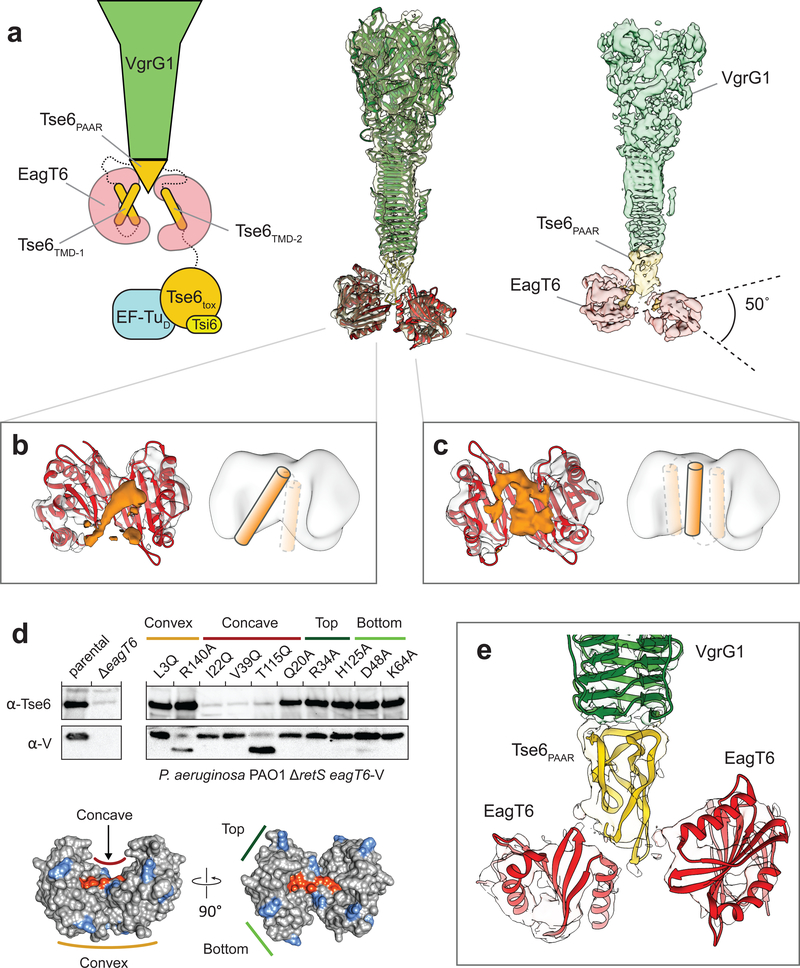
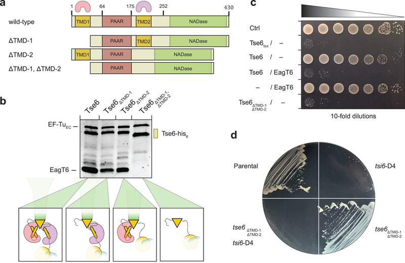
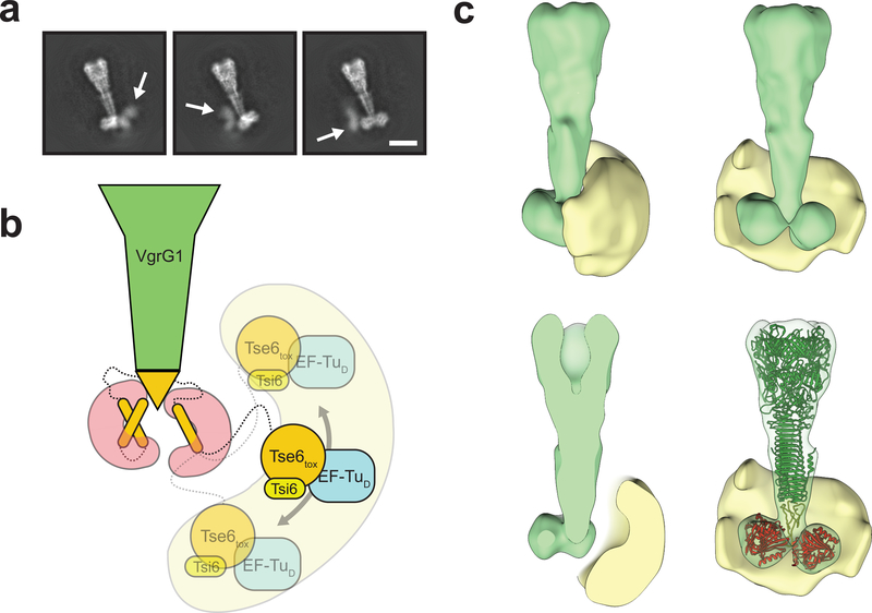
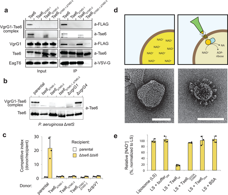
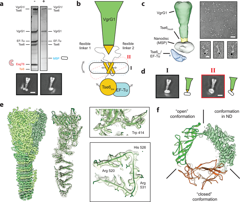
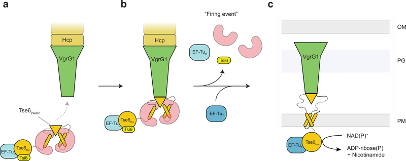
The type VI secretion system (T6SS) primarily functions to mediate antagonistic interactions between contacting bacterial cells, but also mediates interactions with eukaryotic hosts. This molecular machine secretes antibacterial effector proteins by undergoing cycles of extension and contraction; however, how effectors are loaded into the T6SS and subsequently delivered to target bacteria remains poorly understood. Here, using electron cryomicroscopy, we analysed the structures of the Pseudomonas aeruginosa effector Tse6 loaded onto the T6SS spike protein VgrG1 in solution and embedded in lipid nanodiscs. In the absence of membranes, Tse6 stability requires the chaperone EagT6, two dimers of which interact with the hydrophobic transmembrane domains of Tse6. EagT6 is not directly involved in Tse6 delivery but is crucial for its loading onto VgrG1. VgrG1-loaded Tse6 spontaneously enters membranes and its toxin domain translocates across a lipid bilayer, indicating that effector delivery by the T6SS does not require puncturing of the target cell inner membrane by VgrG1. Eag chaperone family members from diverse Proteobacteria are often encoded adjacent to putative toxins with predicted transmembrane domains and we therefore anticipate that our findings will be generalizable to numerous T6SS-exported membrane-associated effectors.


Conflict of interest statement

Competing interests

The authors declare no competing financial interests.

Figures

Figure 1.
Figure 1.. Structure of the “pre-firing” VgrG1-Tse6PAAR-Tse6TMD1,2-EagT62 core complex.
(a) Schematic representation (left), fit of available atomic structures into cryo-EM density map (middle) and color-coded segmented cryo-EM density map (right) of the VgrG1-Tse6-EagT62-EF-Tu-Tsi6 complex. Tse6tox, EF-Tu and Tsi6 are not well resolved due to flexibility; see also Fig. 3. The density corresponding to VgrG1 is shown at a higher threshold for visualization. The exact number of transmembrane helices in each TMD is not known. Predictions give a range from one to three helices (see also Supplementary Fig. 5). Illustrated in the schematic representation are the two most likely arrangements for TMD-2 having either one or three (inset) TMHs. (b,c) Side view on the cavity of the two opposing EagT6 dimers (atomic model in red and density in gray). The additional densities (orange) inside the cavity have a tubular appearance and probably correspond to the transmembrane helices of TMD-1 and TMD-2 of Tse6. In one of the cavities, the enclosed density is less defined indicating a higher degree of flexibility and/or more than one transmembrane helix (c). For corresponding top views, see Supplementary Fig. 1d. Cartoon representations indicate the number of putative transmembrane helices. (d) Single point mutations at the surface of EagT6 identifying residues interacting with Tse6. Western blot analysis of Tse6 and EagT6 levels in the indicated P. aeruginosa strain (upper panel). Different point mutations were introduced in chromosomally VSV-G (V)-tagged EagT6. The lower panel shows the effect of mutated residues mapped on the surface of EagT6: orange - interacting with Tse6, blue - no effect. (e) Close-up view on the interfaces between the two EagT6 dimers and Tse6PAAR. See also Supplementary Figures 1–5 and Supplementary Tables 1–3.
Figure 2.
Figure 2.. EagT6 interacts with both TMDs of Tse6.
(a) Primary domain structure of Tse6 and corresponding TMD-deletion mutants. The two EagT6 dimers are indicated by pink and purple cones to highlight the interaction sites on Tse6. TMD-deletion mutants lack either one TMD (Tse6ΔTMD-1 with Δ1–61 and Tse6ΔTMD-2 with Δ180–222) or both TMDs (Tse6 ΔTMD-1, ΔTMD-2 with Δ1–61 and Δ180–222). (b) Pull-downs of EagT6 and EF-Tu by Tse6wt and TMD-deletion mutants. If one or both of the TMDs are present EagT6 and EF-Tu are pulled down. Absence of both TMDs abolishes EagT6 but not EF-Tu binding. (c) In E. coli toxicity assays, Tse6ΔTMD-1,ΔTMD-2 shows similar toxicity compared to Tse6toxin and the Tse6/EagT6 complex. (d) IPTG-inducible depletion of Tsi6-D4 in P. aeruginosa shows that Tse6ΔTMD-1,ΔTMD-2 is as toxic as Tse6wt. See also Supplementary Figure 6,7 and Supplementary Tables 2 and 3.
Figure 3.
Figure 3.. Tse6tox-EF-Tu-Tsi6 subcomplex is highly flexible.
(a) Representative 2-D class averages showing diffuse densities for the Tse6tox-EF-Tu-Tsi6 subcomplex. White arrows indicate flexible region (see also Supplementary Video 1). Scale bar: 10 nm. (b) Schematic representation of flexibility of the Tse6tox-EF-Tu-Tsi6 subcomplex, which is connected to the more rigid VgrG1-Tse6NTD-EagT62 subcomplex by a linker. (c) Rotated views and cross-sections of filtered 3-D average (green) and 3-D variability (yellow) densities corresponding to the VgrG1-Tse6-EagT62-EF-Tu-Tsi6 complex. The variability density indicates the range of positions taken by the Tse6tox-EF-Tu-Tsi6 subcomplex. Structures of VgrG1 (green), Tse6PAAR (yellow) and EagT6 (red) are fitted in 3-D average volume for orientation. See also Supplementary Tables 2 and 3 and Supplementary Video 1.
Figure 4.
Figure 4.. Crucial role of the TMDs of Tse6 in VgrG1 loading and target cell intoxication.
(a) Immunoprecipitation assay of individual and double TMD deletion mutants in E. coli. Only wild-type Tse6 is able to form the detergent-stable Tse6-VgrG1 complex. EagT6 is VSV-G-tagged, VgrG1 has a FLAG-tag. (b) Western blot analysis of Tse6 levels in the indicated P. aeruginosa strains show that both TMDs are required for high-molecular VgrG1-Tse6 complex formation in vivo. Tse6 only interacts with VgrG1, but not VgrG4, highlighting the specificity of this interaction. (c) Growth competition experiments between P. aeruginosa donor strains and a parental (Δ retS) or Tse6-susceptible (Δ tse6 Δ tsi6) recipient, showing that only wild-type Tse6 has a fitness advantage, while deletion of one or both TMDs is abolishing this effect. Data are means ± standard deviation from three independent biological measurements (n=3). (d) Schematic illustration of NAD+-containing liposomes in the presence and absence of the Tse6-loaded VgrG1 complex with corresponding electron micrographs below. NAD+ is converted into ADP-ribose and nicotinamide (NA). Scale bar: 100 nm. (e) Liposome-based in vitro translocation assay with relative NAD+ levels (normalized to LS), showing degradation of NAD+ after incorporation of the Tse6-loaded VgrG1 complex. DM-containing buffer (bufferDM) showed that the detergent does not cause leakage of liposomes. A catalytically inactive Tse6 mutant (Tse6Q333A,D396A) as well as a Tse6 mutant lacking its C-terminal toxin domain (Tse6Δtox) acted as additional control to exclude perforation of liposomes by the needle-like particles. Data are means ± standard deviation from three independent measurements (n=3). See also Supplementary Figure 8 and Supplementary Tables 2 and 3.
Figure 5.
Figure 5.. The toxin domain of Tse6 spontaneously crosses a lipid bilayer.
(a) Silver stained SDS-PAGE of the Tse6-loaded VgrG1 complex in its “pre-firing” conformation (−) and reconstituted in lipid nanodiscs (+). Upon reconstitution, EagT6 and Tsi6 dissociate from the complex and are exchanged by the nanodisc. Scale bar: 10 nm. (b) Schematic representation of the VgrG1-Tse6-EF-Tu complex reconstituted in lipid nanodiscs. Flexibility caused by (1) lateral movement of the TMDs within the nanodisc, (2) tilting of the nanodisc as well as (3) movement of the Tse6tox-EF-Tu subcomplex impeded structural determination of the bottom part. (c) Low-resolution cryo-EM reconstruction of VgrG1-Tse6-EF-Tu complex embedded in lipid nanodiscs (left) and representative negatively stained electron micrograph areas of the complex (right). Scale bar: 50 nm. Right panel shows three examples of VgrG1-Tse6-EF-Tu complexes in nanodiscs, labeled with 5 nm NTA-coated nanogold to label his-tagged Tse6. Scale bar: 10 nm. (d) Two representative class averages of the VgrG1-Tse6-EF-Tu complex in side view (black I) and tilted view (red II), corresponding to the conformations shown in Fig. 5b. Scale bar: 10 nm. (e) 3.2 Å cryo-EM reconstruction of VgrG1 obtained from the same dataset and applying C3 symmetry. Subunits of trimeric VgrG1 are colored in different green hues (left), fit of atomic model in single subunit (middle), as well as close-ups showing side chain densities (right) of the VgrG1 trimer. (f) Comparison between atomic VgrG1 structures in ‘open’ and ‘closed’ conformations, showing that VgrG1 within the VgrG1-Tse6-EF-Tu complex in nanodiscs adopts an ‘open’ conformation. See also Supplementary Figures 1–3 and 9 and Supplementary Tables 1–3 and Supplementary Video 2.
Figure 6.
Figure 6.. Model for Tse6 effector loading and delivery.
(a) EagT6 (red) binds to the hydrophobic TMDs of Tse6 (orange). This prevents the protein from aggregating and ensures the correct folding of the PAAR domain. The PAAR domain specifically recognizes VgrG1 and mediates the loading of Tse6 onto VgrG1. EagT6 is therefore crucial for the efficient assembly of the Tse6-VgrG1 complex. The binding of EF-Tu (light blue) and Tsi6 (yellow) completes the T6S effector/chaperone complex. (b) Prior to firing EagT6, EF-Tu and Tsi6 dissociate from the complex activating the Tse6toxin. (c) The T6SS punctures the outer membrane of the target cell, forcefully bringing Tse6 into the periplasm. Tse6 spontaneously enters the inner membrane and translocates the Tse6toxin domain across the membrane. On the cytosolic side of the membrane, Tse6toxin binds to EF-Tu and acts as glycohydrolase depleting the cytosolic NAD(P)+ pool. OM – outer membrane, PG – peptidoglycan, IM – inner membrane, D – donor, T – target.

References

    1. Mougous JD et al. A Virulence Locus of Pseudomonas aeruginosa Encodes a Protein Secretion Apparatus. Science 312, 1526–1530 (2006). - PMC - PubMed
    1. Pukatzki S et al. Identification of a conserved bacterial protein secretion system in Vibrio cholerae using the Dictyostelium host model system. PNAS 103, 1528–1533 (2006). - PMC - PubMed
    1. Hood RD et al. A Type VI Secretion System of Pseudomonas aeruginosa Targets a Toxin to Bacteria. Cell Host & Microbe 7, 25–37 (2010). - PMC - PubMed
    1. Russell AB et al. Type VI secretion delivers bacteriolytic effectors to target cells. Nature 475, 343–347 (2011). - PMC - PubMed
    1. Chang YW, Rettberg LA, Ortega DR & Jensen GJ In vivo structures of an intact type VI secretion system revealed by electron cryotomography. EMBO reports e201744072 (2017). doi:10.15252/embr.201744072 - DOI - PMC - PubMed

Publication types

MeSH terms

LinkOut - more resources