. 2018 Sep 3;8(1):13119.
doi: 10.1038/s41598-018-31073-6.

Isoform-specific hyperactivation of calpain-2 occurs presymptomatically at the synapse in Alzheimer's disease mice and correlates with memory deficits in human subjects

Affiliations

Isoform-specific hyperactivation of calpain-2 occurs presymptomatically at the synapse in Alzheimer's disease mice and correlates with memory deficits in human subjects

Faraz Ahmad et al. Sci Rep. .

Abstract

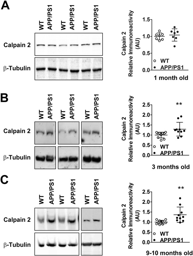
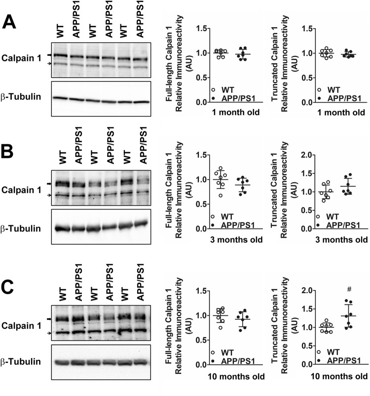
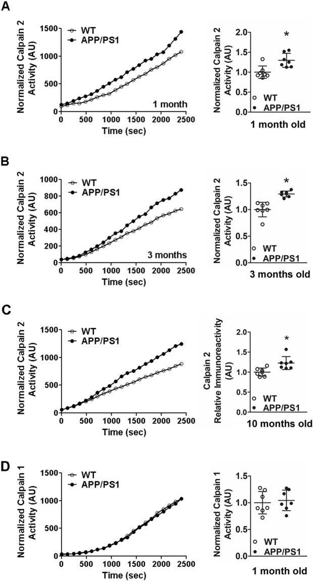
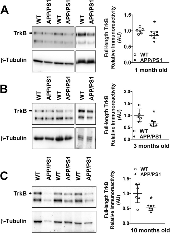
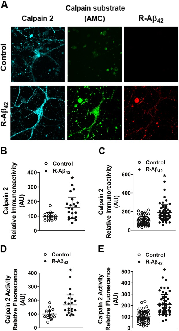
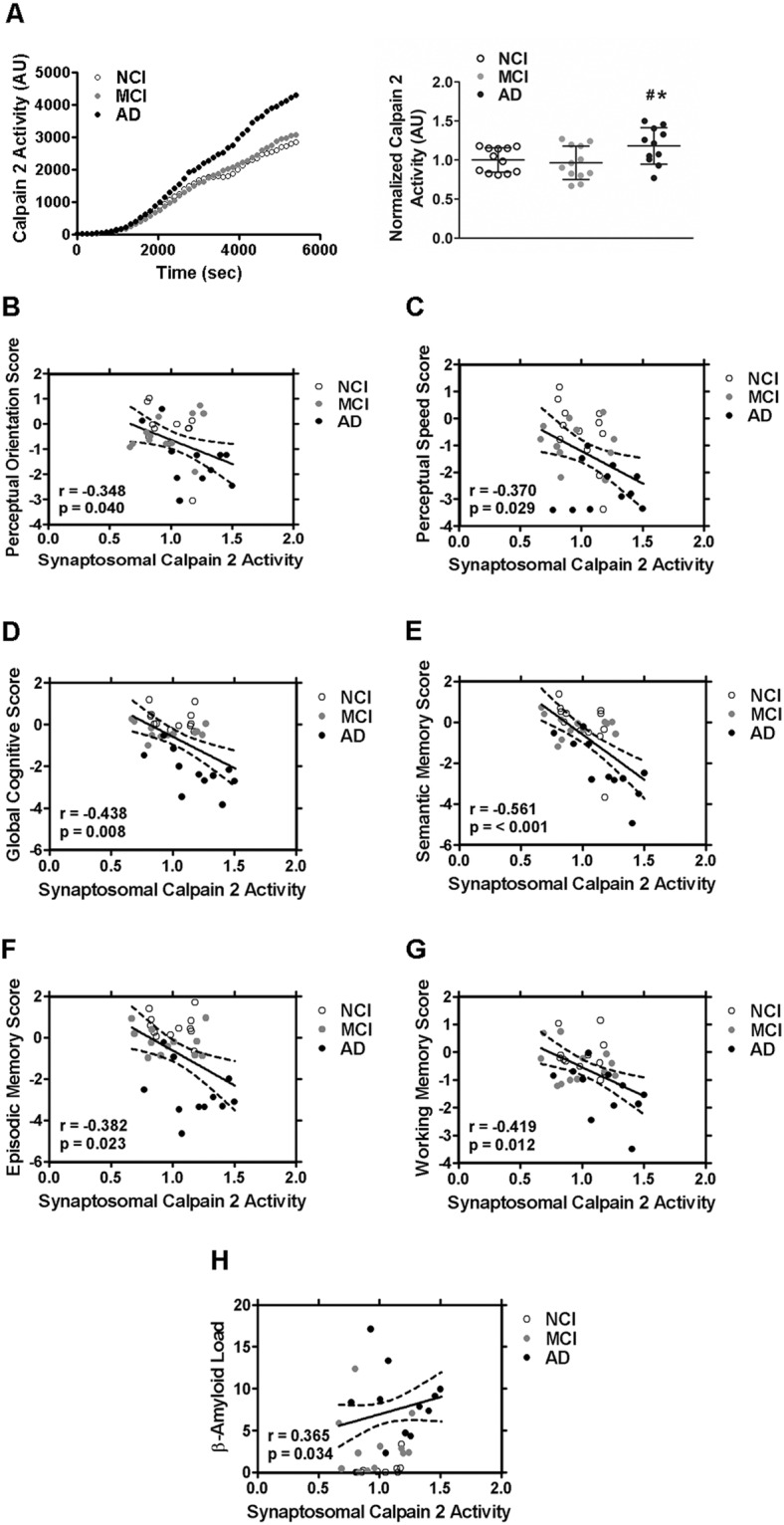
Calpain hyperactivation is implicated in late-stages of neurodegenerative diseases including Alzheimer's disease (AD). However, calpains are also critical for synaptic function and plasticity, and hence memory formation and learning. Since synaptic deficits appear early in AD pathogenesis prior to appearance of overt disease symptoms, we examined if localized dysregulation of calpain-1 and/or 2 contributes to early synaptic dysfunction in AD. Increased activity of synaptosomal calpain-2, but not calpain-1 was observed in presymptomatic 1 month old APPswe/PS1ΔE9 mice (a mouse model of AD) which have no evident pathological or behavioural hallmarks of AD and persisted up to 10 months of age. However, total cellular levels of calpain-2 remained unaffected. Moreover, synaptosomal calpain-2 was hyperactivated in frontal neocortical tissue samples of post-mortem brains of AD-dementia subjects and correlated significantly with decline in tests for cognitive and memory functions, and increase in levels of β-amyloid deposits in brain. We conclude that isoform-specific hyperactivation of calpain-2, but not calpain-1 occurs at the synapse early in the pathogenesis of AD potentially contributing to the deregulation of synaptic signaling in AD. Our findings would be important in paving the way for potential therapeutic strategies for amelioration of cognitive deficits observed in ageing-related dementia disorders like AD.


Conflict of interest statement

The authors declare no competing interests.

Figures

Figure 1
Figure 1
Characterization of synaptosome enrichment and localization of calpain-2 in the synapse. (A) Electron micrograph of synaptosome isolated from mouse brain cortex by sucrose density gradient. Synaptic vesicles in the presynapse (arrow head) and the post synaptic density; PSD (arrow) are marked. Scale bar = 200 nm or 0.2 µm (B) Enrichment of post synaptic density protein (PSD95) in synaptosomes. The synaptosome isolation was performed as described in Materials and Methods. Post nuclear supernatant (PNS) and synaptosomes (Syn; 8 μg of protein from each fraction/lane) were resolved by SDS-PAGE, and immunoblotted against antibodies to PSD95 and tubulin. Upper panel blots were probed for PSD95. Later, these blots were probed for tubulin (lower panel) without stripping. Densitometric analyses for PSD95 levels were normalized to tubulin. Unpaired two-tailed Mann-Whitney test was used. Results are represented as the mean ± SD (n = 6 mice). (C) Colocalization of calpain-2 with homer1 in cortex of WT and APP/PS1 mouse brain. Immunofluorescence staining indicates co-localization of calpain-2 (red) with homer1 (green) in both the cell body and neurites of the neurons. Arrows indicates the immunoreactivity of calpain-2 and homer1 in neurites. Calpain-2 and homer1 co-immunofluorescence was most prominent in neurites as shown in the higher fluorescence micrographs as insets. No staining was seen in mouse cerebral cortex section incubated with IgG control. Scale bar = 75 μm.
Figure 2
Figure 2
1–42 in cortical synaptosomes and in neurons derived from APP/PS1 mice. (A) Total Aβ1–42 levels in synaptosomes prepared from WT and APP/PS1 mice at 3 and 9 months of age were measured using ELISA. Data is represented as mean ± SD (n = 4 mice) and was analyzed using two-tailed Mann Whitney t-test. (B) The products formed following proteolysis of APP in the synaptosomes were detected by immunoblotting with 6E10 antibody and are represented as Aβ monomers, Aβ dimers/β-CTF and full length APP. Synthetic Aβ1–42 was used as standard to confirm the identity of the lower molecular weight bands. ‘*’ indicates a non-specific protein band detected in all samples. (C) Representative confocal images of primary neurons from WT and APP/PS1 mice, immunostained against homer1 (green) and Aβ42 (red) indicates co-localization of Aβ42 with homer1. Scale bar is 20 μm. (D) Representative overlay of high magnification confocal images of primary neurons from WT and APP/PS1 mice - Homer1 (green) and Aβ42 (red). Scale bar is 5 μm. (E) Quantification of Aβ42 signal from soma and neurites of both groups is shown. Data is represented as mean ± SD (n = 6–9 neurons and n = 16–36 neurites from each independent experiment). Mann Whitney two-tailed test was used for statistical analysis.
Figure 3
Figure 3
Protein levels of calpain-2 are increased in synaptosomes of APP/PS1 mice early in the disease pathogenesis. While immunoreactive levels of calpain-2 were unaltered in synaptosomes of 1 month old APP/PS1 mice (A), calpain-2 levels were significantly increased at 3 months (B) and 9–10 months (C) of age when compared to age matched WT controls. Values are mean ± SD (n = 7–10 mice) and ** denotes values significantly different from corresponding controls (p < 0.01; Mann Whitney test). In (A), re-probe for tubulin was performed without stripping and in (B), the blot was cut along the dotted line. Full representative blots are also shown in Supplementary data (Supplementary Figs 3–5).
Figure 4
Figure 4
Protein and activity levels of synaptosomal calpain-1 are unaltered in APP/PS1 mice at presymptomatic ages of 1 and 3 months but truncated synaptic calpain-1 levels are elevated at 10 months. Immunoreactive levels of full-length and truncated calpain-1 (marked as block arrows and thin arrows respectively in representative blots) were unaltered in synaptosomes of 1 month (A) and 3 months (B) old APP/PS1 mice when compared to aged matched WT controls. (C) While full-length synaptosomal calpain-1 levels were unaltered at 10 months, truncated calpain-1 levels were significantly increased. Values are mean ± SD (n = 7 mice) and ‘#’ denotes p = 0.05 (Mann Whitney test). See full representative blots in Supplementary data (Supplementary Figs 6–8).
Figure 5
Figure 5
Global protein levels of calpain-2 are unaltered in PNS of APP/PS1 mice at both pre-symptomatic and early symptomatic ages. Global protein levels of calpain-2 as assessed by immunoreactivity in cortical PNS fractions of APP/PS1 mice were similar to aged matched WT controls at ages of 1 month (A), 3 months (B) and 10 months (C). Values are mean ± SD (n = 7 mice) and p values are calculated using Mann Whitney test. See full representative blots in Supplementary data (Supplementary Figs 9–11).
Figure 6
Figure 6
Activity levels of synaptic calpain-2 are upregulated pre-symptomatically in APP/PS1 mice from 1 month onwards. Immunoprecipitation of calpain-2 was employed to assay isoform-specific activity of calpain-2 in synaptosomal samples isolated from brain cortices of APP/PS1 mice. Increased activity of calpain-2 was observed from 1 month of age (A), persisting up to 3 months (B) and later up to 10 months (C). Isoform specific activity of calpain-1 assayed after immunoprecipitation of calpain-1 (See Supplementary Fig. 1) in cortical synaptosomes of APP/PS1 mice was unchanged at 1 month of age (D). Each of the individual panels show both the representative activity curves and the cumulative data. Values are mean ± SD (n = 7 mice) and * denotes values significantly different from corresponding controls (p < 0.05; Mann Whitney test). See also Supplementary Fig. 1.
Figure 7
Figure 7
Levels of synaptosomal full-length TrkB, a calpain substrate, is reduced in APP/PS1 mice early in the disease pathogenesis. (A) Pre-symptomatic reduction in levels of full-length TrkB, a calpain substrate, was observed in synaptosomes of APP/PS1 mice from 1 month onward. (B) Reduced synaptosomal full-length TrkB levels in cortices of APP/PS1 mice persisted up to 10 months of age. Values are mean ± SD (n = 5–7 mice) and * denotes values significantly different from corresponding controls (p < 0.05; Mann Whitney test). See full representative blots in Supplementary data (Supplementary Figs 12,13).
Figure 8
Figure 8
42 enhances both activity and expression of calpain-2. Primary neurons were exposed to R-Aβ42 for 6 hours and control neurons were untreated. Primary neurons were then exposed to fluorogenic AMC peptide substrate. (A) Representative confocal images are shown, with calpain-2 immunostaining signal in cyan, cleaved AMC peptide signal in green, and R-Aβ42 signal in red. Scale bar is 20 µm. (B,C) Increased expression of calpain-2 levels was observed in both soma and neurites of neurons treated with R-Aβ42 compared to control neurons. Increase with calpain-2 levels was associated with a concomitant increase in its activity, as assessed by generation of fluorescent peptide upon cleavage of AMC peptide; both in soma and neurites of primary neurons pre-treated with R-Aβ42 (D,E). Data is represented as mean ± SD (n = 16–18 neurons) and * indicates statistical significance (p < 0.05; Mann Whitney test).
Figure 9
Figure 9
Protein levels of calpain-2 are higher in frontal neocortical tissue samples of post-mortem brains of AD patients in a synaptosome-specific manner. (A) Immunoreactive levels of calpain-2 were increased in synaptosomes isolated from post-mortem brains of AD patients when compared to subjects with MCI as well as subjects with NCI. (B) Global calpain-2 protein levels were unaltered in post-mortem AD brains. Values are mean ± SD (n = 12 subjects) and * and # denote significant difference from NCI and MCI groups respectively (p < 0.05; ANOVA followed by post hoc tests with Newman-Keuls correction). See full representative blots in Supplementary data (Supplementary Figs 14,15).
Figure 10
Figure 10
Activity levels of calpain-2 are elevated in synaptosomes isolated from frontal neocortical tissue samples of post-mortem brains from AD patients. (A) Activity of synaptosomal calpain-2 immunoprecipitated from post-mortem brains of subjects with AD dementia and was found to be significantly higher compared to subjects with no cognitive impairment (NCI) or mild cognitive impairment (MCI). A significant negative correlation was observed between synaptosomal calpain-2 activity and performance of the subjects in tests for perceptual orientation (B) and speed (C), global cognition (D), semantic memory (E), episodic working (F) and as well as working memory (G) tests. Increase in calpain-2 activity in synaptosomal samples from post-mortem human brains also correlated with increase in amyloid deposits in the brain (H). Values are mean ± SD (n = 12 subjects) and * and # denote significant difference from NCI and MCI groups respectively (p < 0.05; ANOVA followed by post hoc tests with Newman-Keuls correction). See also Supplementary Fig. 2.

Similar articles

Cited by

References

    1. Lepeta K, et al. Synaptopathies: synaptic dysfunction in neurological disorders. J Neurochem. 2016;138:785–805. doi: 10.1111/jnc.13713. - DOI - PMC - PubMed
    1. Wang Z, Yang L, Zheng H. Role of APP and Aβ in synaptic physiology. Curr Alzheimer Res. 2012;9:217–226. doi: 10.2174/156720512799361691. - DOI - PubMed
    1. Saido TC, Sorimachi H, Suzuki K. Calpain: new perspectives in molecular diversity and physiological-pathological involvement. FASEB J. 1994;8:814–822. doi: 10.1096/fasebj.8.11.8070630. - DOI - PubMed
    1. Baudry M, Chou MM, Bi X. Targeting calpain in synaptic plasticity. Expert Opin Ther Targets. 2013;17:579–592. doi: 10.1517/14728222.2013.766169. - DOI - PMC - PubMed
    1. Camins A, Verdaguer E, Folch J, Pallàs M. Involvement of calpain activation in neurodegenerative processes. CNS Drug Rev. 2006;12:135–148. doi: 10.1111/j.1527-3458.2006.00135.x. - DOI - PMC - PubMed

Publication types

MeSH terms