. 2018 Sep 3;8(1):13146.
doi: 10.1038/s41598-018-31567-3.

Inhibiting Multiple Deubiquitinases to Reduce Androgen Receptor Expression in Prostate Cancer Cells

Affiliations

Inhibiting Multiple Deubiquitinases to Reduce Androgen Receptor Expression in Prostate Cancer Cells

Alicia de Las Pozas et al. Sci Rep. .

Abstract

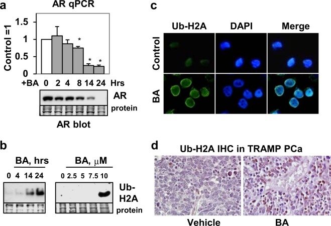
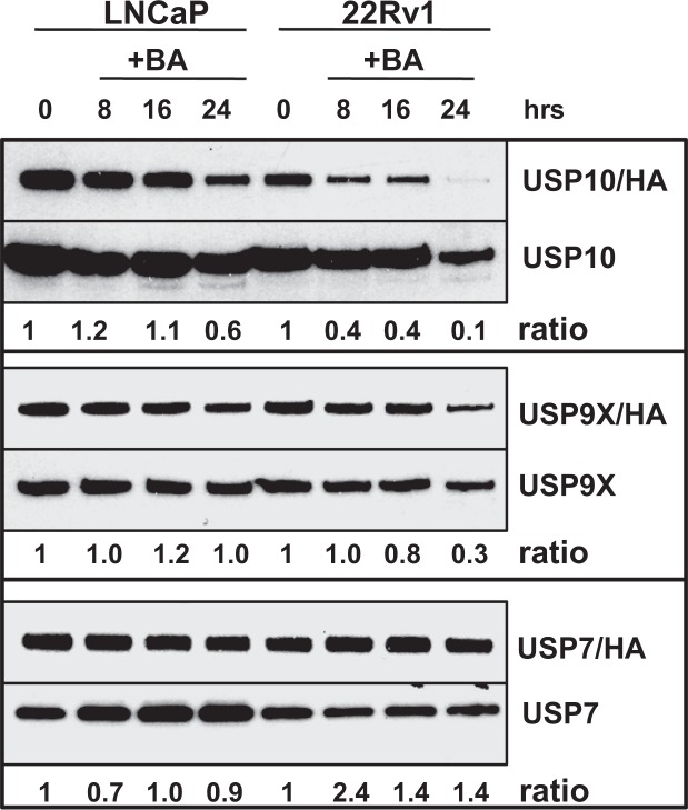
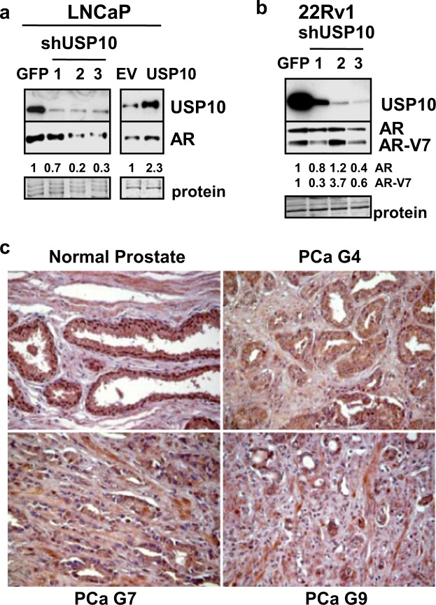
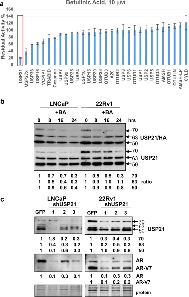
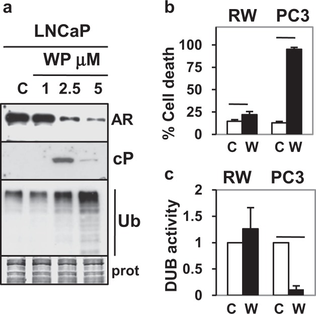
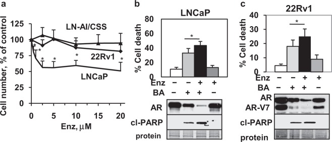
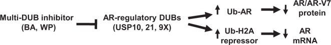
Prostate cancer (PCa), a leading cause of cancer-related death in men, becomes resistant to androgen deprivation therapy by inducing androgen receptor (AR) activity, which is known as castration-resistant PCa (CRPC). Enzalutamide is an approved drug that inhibits AR activity and increases overall survival. However, resistance to enzalutamide develops rapidly often by increasing AR activity, suggesting that new therapies are required for CRPC. We investigated whether betulinic acid (BA), a small molecule from plants that inhibits multiple deubiquitinases (DUBs), reduces AR, and selectively kills PCa cells, can provide an adjuvant strategy for CRPC. Our data indicated that BA reduced AR protein stability and mRNA expression, making it an attractive agent for CRPC. BA decreased AR mRNA possibly by inhibiting a histone 2A DUB thereby increasing ubiquitinated histone 2A, a transcriptional repressor. We identified multiple and specific DUBs inhibited by BA either in PCa cells or using recombinant DUBs. Similar results were obtained using another multi-DUB inhibitor WP1130, suggesting that these DUB inhibitors can decrease AR expression and increase PCa-specific death. Our results also suggest that combining multi-DUB inhibitors BA or WP1130 with enzalutamide may provide a novel strategy for CRPC by further decreasing AR expression and increasing apoptotic cell death.


Conflict of interest statement

The authors declare no competing interests.

Figures

Figure 1
Figure 1
BA increases apoptotic and necrotic cell death in multiple PCa cell lines. Trypan blue exclusion assay (LNCaP 48 h and DU145, PC3 72 h) showed that CsA (10 μM; inhibits necrosis) reduced BA (10 μM) cell death only in PC3. Pan-caspase inhibitor QVD (10 μM; inhibits apoptosis) reduced BA cell death and cl-PARP (western blot) in LNCaP and DU145. QVD also reduced cl-PARP in PC3 but had no effect on BA cell death. Addition of CsA and QVD to BA reduced cell death in LNCaP, DU145, and PC3. Coomassie blue stain of total protein transferred to the membrane was the loading control for western blot. Blot images were cropped for clarity of the presentation. *P < 6 × 10−3; n = 6, 3 independent experiments.
Figure 2
Figure 2
BA decreases AR variant associated with Enz resistance and lowers AR protein stability. (a) Western blot showed that BA (10 μM) decreased AR and AR-V7 variant and increased cl-PARP, poly-Ub without affecting actin in 22Rv1 cells. (b) Western blot showed that Chx (25 μg/ml) + BA (Chx/BA) decreased AR and AR-V7 protein more rapidly especially at 8, 16, and 24 h compared to Chx + control (Chx/C) in 22Rv1 cells. Actin was unaffected. Loading control (protein). Blot images were cropped for clarity of the presentation.
Figure 3
Figure 3
BA increases Ub-AR in LNCaP and 22Rv1. (a) Western blot showed that BA (10 μM; 20 h) increased Ub-AR (~200 kDa) compared to control in LNCaP after immunoprecipitation (IP) with AR and immunoblot (IB) with Ub. AR IP and AR IB showed the expected signal for AR. Negative control IP with mouse Ig showed no signal. Input lysate (Lys), shown to the left, was run on a separate gel. (b) Ub-AR also increased in BA compared to control LNCaP after Ub IP and AR IB. Ub IP and Ub IB showed the expected signal compared to negative control IP with mouse Ig. Total protein (Lys) was run on the same gel. Input Lys, shown to the left, was run on a separate gel. (c) In 22Rv1, Ub IP and AR IB also increased Ub-AR in BA compared to control. Ub IP and Ub IB showed the expected signal compared to negative control IP with mouse Ig. Total protein (Lys) was run on the same gel (exposure time for AR was less than IP samples). Input Lys, shown to the left, was run on a separate gel. Vertical line in (a,c) indicated samples were from the same blot but not in sequence. Molecular weight markers in kDa are shown to the left of IP/IB blots. Blot images were cropped for clarity of the presentation.
Figure 4
Figure 4
UPS but not AKT, ERK, or JNK inhibition blocks BA decrease of AR in LNCaP. Trypan blue and western blot showed that the (a) UPS inhibitor Btz (5 nM, 24 h) antagonized BA decrease in AR and increase in cell death, cl-PARP whereas (b) AKT inhibitor LY294002 (20 μM, 24 h), (c) ERK inhibitor U0126 (10 μM, 24 h), and (d) JNK inhibitor SP600125 (20 μM, 48 h) did not influence the BA decrease in AR or antagonize cell death/cl-PARP. AKT and ERK but not JNK inhibition further increased BA cell death. Activity of inhibitors was shown by blocking the BA increase in P-AKT, P-ERK, and P-JNK. There were no changes in total ERK or JNK; AKT decreased with BA. *P < 0.03); n = 6, 3 independent experiments. Loading control (protein). Blot images were cropped for clarity of the presentation.
Figure 5
Figure 5
BA increases Ub-H2A transcriptional repressor and decreases AR mRNA. (a) qPCR showed that BA reduced AR mRNA (*P < 0.002; n = 4, 2 independent experiments) and protein (western blot) starting at 8 h in LNCaP. (b) Western blot in LNCaP showed that BA increased Ub-H2A (23 kDa) at 14 and 24 h (left blot); only the 10 μM concentration of BA known to inhibit DUB activity was able to increase Ub-H2A (24 h; right blot). Loading control (protein). Blot images in (a,b) were cropped for clarity of the presentation. (c) Immunofluorescence in LNCaP showed that BA (24 h) increased Ub-H2A (green) in the nucleus (DAPI stain, blue) greater than control cells. (d) IHC of PCa showed greater nuclear Ub-H2A (x200, dark brown color) in BA compared to vehicle control treated TRAMP mice.
Figure 6
Figure 6
BA inhibits USP10 in LNCaP and 22Rv1. Western blot analysis of DUB labeling assay using HA-UbVS and USP10, 9X, and 7 specific antibodies. Results showed that BA inhibited USP10 activity (ratio of USP10/HA/total USP10 values shown below; 0 h = 1). BA inhibited USP9X activity in 22Rv1 but not in LNCaP. There was little effect of BA on USP7 activity. Blot images were cropped for clarity of the presentation. Similar results were obtained in an additional experiment.
Figure 7
Figure 7
USP10, a candidate AR-regulatory DUB inhibited by BA, is variably expressed in human PCa tissues. (a) Western blot showed that stable knockdown (5- to 14-fold) of USP10 in LNCaP with 3 different shRNAs (shUSP10-1, -2, -3) reduced AR protein by 1.5- to 5-fold compared to shGFP control. Stable overexpression of USP10 (6-fold) increased AR protein 2-fold compared to empty vector (EV) control. Loading control (protein). (b) In 22Rv1, stable knockdown of USP10 (6- to 100-fold) reduced AR and AR-V7 protein by 1.5- to 3-fold in 2 of 3 shRNAs. Blot images in (a,b) were cropped for clarity of the presentation. (c) Representative IHC images of USP10 expression (x200, dark brown color) in human PCa tissues compared to normal prostate using a tissue microarray. Results showed that USP10 was highly expressed in cytoplasm and nucleus of epithelial cells in normal prostate. With higher Gleason (G7 and 9 compared to G4) grades of PCa, USP10 was more variably expressed with less nuclear localization.
Figure 8
Figure 8
USP21 is a novel AR-regulatory DUB inhibited by BA. (a) MALDI-TOF analysis showed that 10 μM BA inhibited recUSP21 activity by 80% (red rectangle). BA also inhibited recUSP27 (60%), USP36 (40%), and USP16 (40%) to a lesser extent compared to USP21. Unlike results in LNCaP and 22Rv1 PCa cells (Fig. 6), BA had little effect on recUSP10 or 9X. (b) Western blot analysis of DUB labeling assay in LNCaP and 22Rv1 using HA-UbVS and USP21 specific antibody. Results showed that in LNCaP, BA inhibited 3 isoforms (70, 63, and 50 kDa) of USP21 activity (ratio of USP21/HA/total USP21 values for 3 isoforms shown below; 0 h = 1). In 22Rv1, BA inhibited the 70 kDa but not the 63 or 50 kDa isoforms of USP21. Blot images were cropped for clarity of the presentation. Similar results were obtained in an additional experiment. (c) Western blot showed that stable partial knockdown (2- to 10-fold) of USP21 isoforms in LNCaP and 22Rv1 with 3 different shRNAs (shUSP21-1, -2, -3) reduced AR and AR-V7 protein by 3- to 10-fold compared to shGFP controls. Loading control (protein). Blot images were cropped for clarity of the presentation.
Figure 9
Figure 9
WP DUB inhibitor also decreases AR protein and has little effect in non-cancer cells. (a) Western blot analysis showed that WP (2.5 and 5 but not 1 μM; 16 h) decreased AR protein and increased cl-PARP (cP), poly-Ub in LNCaP. Loading control (prot). Blot images were cropped for clarity of the presentation. (b) Trypan blue assay showed that WP (W; 5 μM, 24 h) only slightly increased cell death in non-cancer RWPE-1 (RW) prostate epithelial cells compared to vehicle control (C) (P = 0.006) but greatly increased cell death in PC3 (P = 4 × 10−9). (c) DUB assay showed that WP (W; 5 μM, 24 h) did not inhibit DUB activity in RW but greatly inhibited DUB activity in PC3 (P = 0.002). In (b,c), n = 4–6, 2 independent experiments.
Figure 10
Figure 10
BA + Enz is a novel combination that decreases AR and increases cell death in PCa cells. (a) Cell viability assay (72 h) showed that LNCaP PCa but not LN-AI/CSS or 22Rv1 CRPC cells were sensitive to Enz (*P < 0.004; n = 9, 3 independent experiments). (b,c) Trypan blue exclusion (LNCaP, 16 h; 22Rv1, 24 h) and western blot (LNCaP, 24 h; 22Rv1, 16 h cl-PARP, 24 h AR) showed that BA (10 μM) + Enz (10 μM) further decreased AR and increased cell death and cl-PARP (cP) in LNCaP and 22Rv1 compared to BA alone (*P < 0.02; n = 6–9, 3 independent experiments). Loading control (protein). Blot images were cropped for clarity of the presentation.
Figure 11
Figure 11
BA inhibits specific AR-regulatory DUBs to decrease AR protein and mRNA. Multi-DUB inhibitors BA and WP are proposed to inhibit AR-regulatory DUBs USP10, 21, and 9X in PCa cells to (1) increase poly Ub and Ub-AR/protein degradation and decrease AR/AR-V7 protein and (2) increase Ub-H2A repressor and decrease AR transcription (mRNA).

References

    1. Perner S, et al. Adaptive responses of androgen receptor signaling in castration-resistant prostate cancer. Oncotarget. 2015;6:35542–35555. doi: 10.18632/oncotarget.4689. - DOI - PMC - PubMed
    1. Attard G, et al. Prostate cancer. Lancet. 2016;387:70–82. doi: 10.1016/S0140-6736(14)61947-4. - DOI - PubMed
    1. Chen CD, et al. Molecular determinants of resistance to antiandrogen therapy. Nat Med. 2004;10:33–39. doi: 10.1038/nm972. - DOI - PubMed
    1. Scher HI, et al. Increased survival with enzalutamide in prostate cancer after chemotherapy. N Engl J Med. 2012;367:1187–1197. doi: 10.1056/NEJMoa1207506. - DOI - PubMed
    1. Scher HI, et al. Antitumour activity of MDV3100 in castration-resistant prostate cancer: a phase 1–2 study. Lancet. 2010;375:1437–1446. doi: 10.1016/S0140-6736(10)60172-9. - DOI - PMC - PubMed

Publication types

MeSH terms