Review
. 2018 Aug 20:12:268.
doi: 10.3389/fncel.2018.00268. eCollection 2018.

Interactions Between Neural Progenitor Cells and Microglia in the Subventricular Zone: Physiological Implications in the Neurogenic Niche and After Implantation in the Injured Brain

Affiliations
Review

Interactions Between Neural Progenitor Cells and Microglia in the Subventricular Zone: Physiological Implications in the Neurogenic Niche and After Implantation in the Injured Brain

Esperanza R Matarredona et al. Front Cell Neurosci. .

Abstract

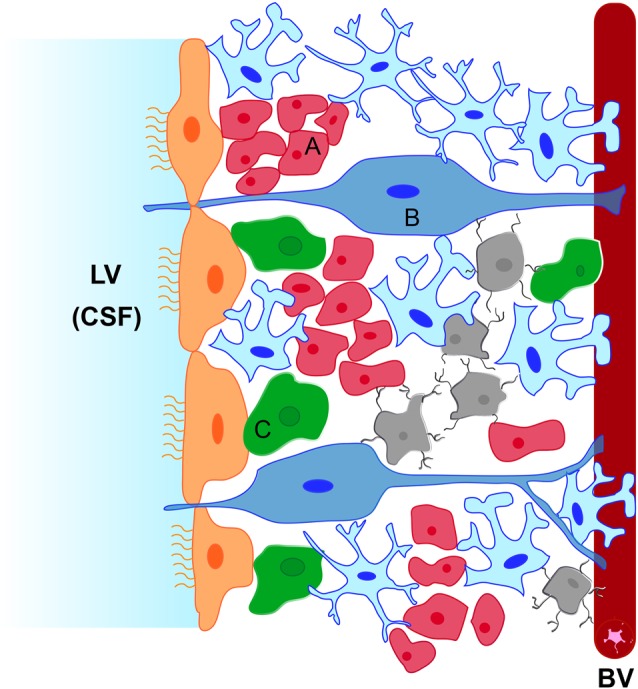
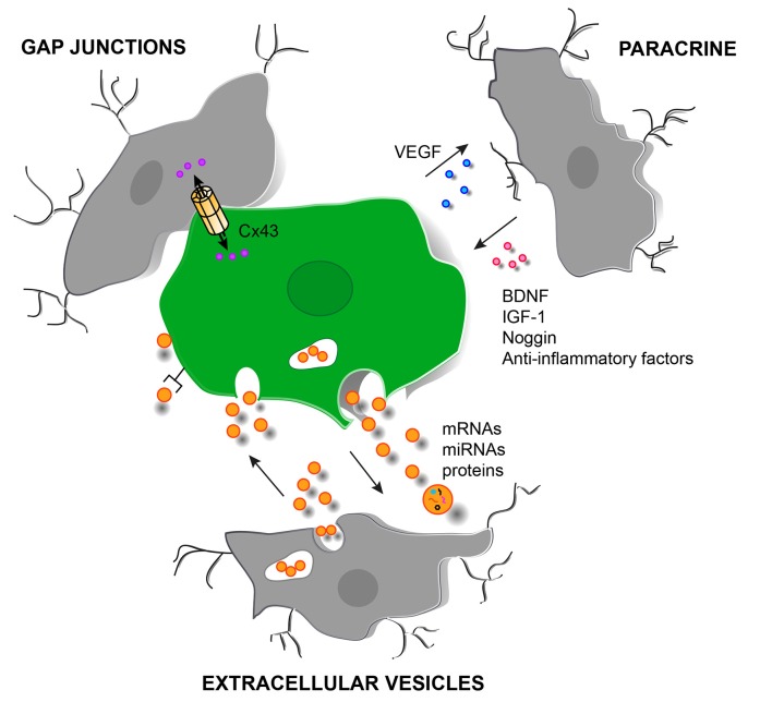
The adult subventricular zone (SVZ) of the mammalian brain contains neural progenitor cells (NPCs) that continuously produce neuroblasts throughout life. These neuroblasts migrate towards the olfactory bulb where they differentiate into local interneurons. The neurogenic niche of the SVZ includes, in addition to NPCs and neuroblasts, astrocytes, ependymal cells, blood vessels and the molecules released by these cell types. In the last few years, microglial cells have also been included as a key component of the SVZ neurogenic niche. Microglia in the SVZ display unique phenotypic features, and are more densely populated and activated than in non-neurogenic regions. In this article we will review literature reporting microglia-NPC interactions in the SVZ and the role of this bilateral communication in microglial function and in NPC biology. This interaction can take place through the release of soluble factors, extracellular vesicles or gap junctional communication. In addition, as NPCs are used for cell replacement therapies, they can establish therapeutically relevant crosstalks with host microglia which will also be summarized throughout the article.

Keywords: extracellular vesicles; gap junctions; microglia; neural progenitor cells; neurogenic niche; paracrine communication; subventricular zone.


Figures

Figure 1
Figure 1
Schematic representation of the subventricular zone (SVZ) neurogenic niche. The niche is located underneath the ependymal cells (in orange) lining the lateral ventricle (LV). It is constituted by type B neural stem cells (in blue, B) which can activate and generate type C neural progenitor cells (NPCs; in green, C) that proliferate rapidly and generate type A neuroblasts (in red, A). Type B cells extend a short apical process to contact the cerebrospinal fluid (CSF) and a long basal process that terminate on blood vessels (BVs). Astrocytes (in pale blue) contact BVs, ependymal cells and type C cells and surround migrating neuroblasts towards the olfactory bulb. Microglial cells (in gray) contact type A, type B and type C cells and also BVs.
Figure 2
Figure 2
Schematic representation of different cell signaling pathways involved in the interaction between grafted NPCs and host microglia in the injured brain. Paracrine signaling with the release of soluble factors from SVZ-derived NPCs (in green) and microglia (in gray). Direct molecule interchange mediated by gap junctional communication can also occur between microglia/macrophages and grafted NPCs. In addition, extracellular vesicles (orange circles) can be released by NPCs and by microglia with the possibility to deliver bioactive molecules such as mRNAs, miRNAs and proteins. The delivery can be carried out in different ways: (i) by endocytosis of the vesicle; (ii) by activation of surface receptors; and (iii) by membrane fusion or by a Cx43-dependent mechanism. Abbreviations: BDNF, brain derived-neurotrophic factor; Cx43, connexin 43; IGF-1, insulin-like growth factor 1; VEGF, vascular endothelial growth factor.

References

    1. Aarum J., Sandberg K., Haeberlein S. L. B., Persson M. A. A. (2003). Migration and differentiation of neural precursor cells can be directed by microglia. Proc. Natl. Acad. Sci. U S A 100, 15983–15988. 10.1073/pnas.2237050100 - DOI - PMC - PubMed
    1. Åkerblom M., Sachdeva R., Barde I., Verp S., Gentner B. (2015). MicroRNA-124 is a subventricular zone neuronal fate determinant. J. Neurosci. 32, 8879–8889. 10.1523/JNEUROSCI.0558-12.2012 - DOI - PMC - PubMed
    1. Ali I., Chugh D., Ekdahl C. T. (2015). Role of fractalkine-CX3CR1 pathway in seizure-induced microglial activation, neurodegeneration, and neuroblast production in the adult rat brain. Neurobiol. Dis. 74, 194–203. 10.1016/j.nbd.2014.11.009 - DOI - PubMed
    1. Alvarez-Buylla A., García-Verdugo J. M. (2002). Neurogenesis in adult subventricular zone. J. Neurosci. 22, 629–634. 10.1523/JNEUROSCI.22-03-00629.2002 - DOI - PMC - PubMed
    1. Antonucci F., Turola E., Riganti L., Caleo M., Gabrielli M., Perrotta C., et al. . (2012). Microvesicles released from microglia stimulate synaptic activity via enhanced sphingolipid metabolism. EMBO J. 31, 1231–1240. 10.1038/emboj.2011.489 - DOI - PMC - PubMed

LinkOut - more resources