Review
. 2018 Aug 20:9:682.
doi: 10.3389/fneur.2018.00682. eCollection 2018.

Research Progress in Understanding the Relationship Between Heme Oxygenase-1 and Intracerebral Hemorrhage

Affiliations
Review

Research Progress in Understanding the Relationship Between Heme Oxygenase-1 and Intracerebral Hemorrhage

Qian-Qian Li et al. Front Neurol. .

Abstract

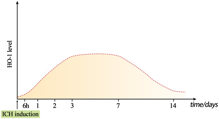
Intracerebral hemorrhage (ICH) is a fatal acute cerebrovascular disease, with a high morbidity and mortality. Following ICH, erythrocytes release heme and several of its metabolites, thereby contributing to brain edema and secondary brain damage. Heme oxygenase is the initial and rate-limiting enzyme of heme catabolism, and the expression of heme oxygenase-1 (HO-1) is rapidly induced following acute brain injury. As HO-1 exerts it effects via various metabolites, its role during ICH remains complex. Therefore, in-depth studies regarding the role of HO-1 in secondary brain damage following ICH may provide a theoretical basis for neuroprotective function after ICH. The present review aims to summarize recent key studies regarding the effects of HO-1 following ICH, as well as its influence on ICH prognosis.

Keywords: heat shock protein 32; heme; heme oxygenase-1; intracerebral hemorrhage; microglia; neurological impairment.


Figures

Figure 1
Figure 1
Dynamic changes in HO-1 levels after intracerebral hemorrhage (ICH). Under normal conditions, HO-1 content in the brain is very low. The expression of HO-1 can be rapidly induced approximately 6 h after ICH, and reaches a peak at days 3–7. The level of HO-1 may gradually decrease on day 7, but it will remain present for at least 14 days.
Figure 2
Figure 2
The expression of HO-1 in microglia and astrocytes after intracerebral hemorrhage (ICH). HO-1 is expressed in both microglia and astrocytes after ICH. HO-1 is mainly expressed in microglia in the early stage of ICH; it is primarily expressed in astrocytes in the late stage of ICH. Microglia can be polarized to two phenotypes, M1 and M2, which are in dynamic flux during ICH. After ICH, M1 microglia can produce pro-inflammatory factors, leading to destruction of the blood-brain barrier, brain edema, white matter damage, iron accumulation, and neurological deficits; M2 microglia can produce anti-inflammatory factors and promote hematoma clearance through phagocytosis and promotion of angiogenesis. In the late stage of ICH, high level HO-1 expression in astrocytes may contribute to increased cell viability, maintenance of blood-brain barrier integrity, and reduction of mortality.
Figure 3
Figure 3
The products of heme metabolism catalyzed by HO-1 (heme oxygenase (HO-1) metabolizes heme). After intracerebral hemorrhage (ICH), HO-1 can catalyze heme to produce biliverdin, carbon monoxide (CO), and free iron. Iron can produce ferritin to exert cytoprotection and anti-oxidative effects; it can also be oxidized to Fe3+, which contributes to brain edema, oxidative stress, neuronal death, blood-brain barrier damage, and brain atrophy. CO can play anti-apoptotic, anti-inflammatory, anti-proliferative, and vasodilating effects through the sGC/cGMP and p38MAPK pathways. Biliverdin is rapidly converted to bilirubin under the BVR, which has anti-inflammatory, anti-oxidative and anti-proliferative effects.
Figure 4
Figure 4
Schematic representation of cellular protection mechanism conferred by Nrf2-ARE pathway after intracerebral hemorrhage (ICH). Under normal conditions, Nrf2 forms a complex with actin-bound Keap1 and Cullin 3, which are isolated in the cytoplasm. Heme released after ICH, along with LPS and oxidative stress, can induce reactive oxygen species (ROS), which can induce and mediate dissociation of the Nrf2-Keap1 complex. Then, Nrf2 translocates to the nucleus and heterodimerizes with Maf. Subsequently, Nrf2 binds to antioxidant response elements (ARE) in the promoter regions of HO-1 and other target genes, activating transcription. Bach1 is an inhibitor of the Nrf2-ARE signaling pathway; thus, the removal of Bach1 is a prerequisite for transcription of this pathway.

Similar articles

Cited by

References

    1. Kim JY, Bae HJ. Spontaneous intracerebral hemorrhage: management. J Stroke (2017) 19:28–39. 10.5853/jos.2016.01935 - DOI - PMC - PubMed
    1. Steiner T, Al-Shahi Salman R, Beer R, Christensen H, Cordonnier C, Csiba L, et al. . European Stroke Organisation (ESO) guidelines for the management of spontaneous intracerebral hemorrhage. Int J Stroke (2014) 9:840–55. 10.1111/ijs.12309 - DOI - PubMed
    1. Donnan GA, Hankey GJ, Davis SM. Intracerebral haemorrhage: a need for more data and new research directions. Lancet Neurol. (2010) 9:167–76. 10.1016/S1474-4422(10)70001-6 - DOI - PubMed
    1. Feigin VL, Lawes CM, Bennett DA, Barker-Collo SL, Parag V. Worldwide stroke incidence and early case fatality reported in 56 population-based studies: a systematic review. Lancet Neurol. (2009) 8:355–69. 10.1016/S1474-4422(09)70025-0 - DOI - PubMed
    1. Li Y, Yang R, Li Z, Tian B, Zhang X, Wang J, et al. . Urokinase vs. tissue-type plasminogen activator for thrombolytic evacuation of spontaneous intracerebral hemorrhage in basal ganglia. Front Neurol. (2017) 8:371. 10.3389/fneur.2017.00371 - DOI - PMC - PubMed