Case Reports
. 2018 Dec;39(12):1995-2007.
doi: 10.1002/humu.23639. Epub 2018 Sep 17.

Insights into the genotype-phenotype correlation and molecular function of SLC25A46

Affiliations
Case Reports

Insights into the genotype-phenotype correlation and molecular function of SLC25A46

Alexander J Abrams et al. Hum Mutat. 2018 Dec.

Abstract

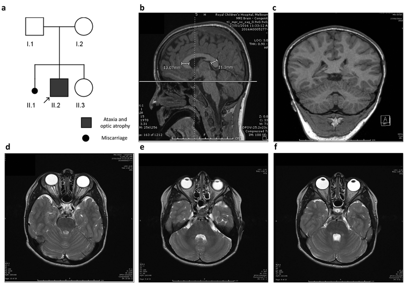
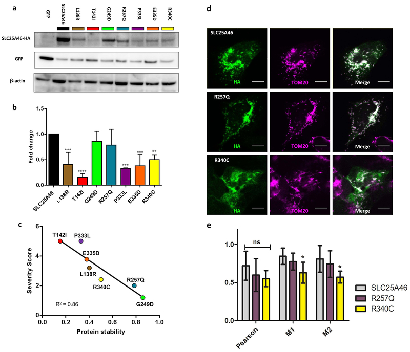
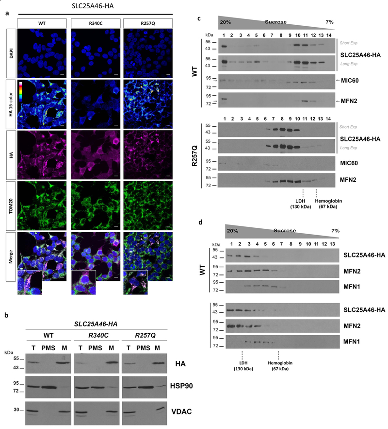
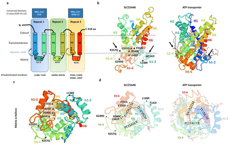
Recessive SLC25A46 mutations cause a spectrum of neurodegenerative disorders with optic atrophy as a core feature. We report a patient with optic atrophy, peripheral neuropathy, ataxia, but not cerebellar atrophy, who is on the mildest end of the phenotypic spectrum. By studying seven different nontruncating mutations, we found that the stability of the SLC25A46 protein inversely correlates with the severity of the disease and the patient's variant does not markedly destabilize the protein. SLC25A46 belongs to the mitochondrial transporter family, but it is not known to have transport function. Apart from this possible function, SLC25A46 forms molecular complexes with proteins involved in mitochondrial dynamics and cristae remodeling. We demonstrate that the patient's mutation directly affects the SLC25A46 interaction with MIC60. Furthermore, we mapped all of the reported substitutions in the protein onto a 3D model and found that half of them fall outside of the signature carrier motifs associated with transport function. We thus suggest that there are two distinct molecular mechanisms in SLC25A46-associated pathogenesis, one that destabilizes the protein while the other alters the molecular interactions of the protein. These results have the potential to inform clinical prognosis of such patients and indicate a pathway to drug target development.

Keywords: Ataxia; Mitochondria; Optic atrophy; SLC25A46.


Conflict of interest statement

Conflict of Interest Statement

The Authors have no conflicts of interest to disclose.

Figures

Figure 1
Figure 1. Pedigree of SLC25A46 patient and MRI images.
(a) Pedigree of index patient (arrow) homozygous for the c.770G>A p.Arg257Gln (p.R257Q) variant. Both parents were confirmed carriers for the mutation. (b-f) Sagittal, coronal, and serial axial MRI images obtain at the age of 5½ years, demonstrating normal appearance of the cerebellar vermis and hemispheres. The corpus callosum is diffusely thick with the genu slightly thicker than the splenium (b). The cerebellum and optic nerve appearance are within the normal limits for age.
Figure 2
Figure 2. Genomic positions and Clustal alignments of reported mutations in SLC25A46.
(a) Schematic gene diagram of SLC25A46 (NM_138773.1, 1,257 base pairs, 418 amino acids). The relative locations of mutations are indicated by red arrows with the letters correspond to the family ID’s listed in (table 1), new patient is A*. The mitochondrial solute carrier domains (MC) are depicted in blue. (b) Clustal alignment demonstrating the conservation of residues in human, cow, mouse, zebrafish, nematode, and the two paralogues in drosophila.
Figure 3
Figure 3. Correlation of protein levels, severity of patient symptoms, and mitochondrial targeting of the SLC25A46 substitution variants.
(a) Representative Western blot of transiently transfected HEK293T cells with SLC25A46-HA mutations and GFP. Blots were probed against HA, GFP, and β-actin. The HA signal was normalized to GFP to control for the transfection efficiency. (b) Histogram shows the mean ± SD from three independent experiments, the relative protein levels of the mutations were normalized to SLC25A46 WT for each experiment. All of the mutations are significantly reduced in comparison to wildtype SLC25A46 with the exception of p.R257Q and p.G249D which were trending but not significant. (c) Correlation plot between the clinical severity scores from (Table 2) and the average relative protein levels determined by the Western blots. (d) Colocalization of wildtype SLC25A46, p.R257Q, and p.R340C with the mitochondria outer membrane marker TOM20 in U2OS cells. Wildtype and p.R257Q colocalize strongly with TOM20, while p.R340C shows less colocalization with the mitochondria. Scale bar 10 μm. (e) Quantification of three measures of colocalization; Pearson, Meanders 1 (M1), and Meanders 2 (M2). Significance was determined by a 2-way ANOVA with Bonferroni posttest with 6 cells per condition.
Figure 4
Figure 4. Analysis of SLC25A46 expression and native molecular weight in stable transfected HEK293T cells.
(a) Immunocytochemistry of cells expressing wildtype (WT), p.R257Q, or p.R340C variants. DAPI was used to visualize nuclei and an anti-TOM20 antibody was used as mitochondrial marker (b) Cell fractionation analysis of wildtype (WT), p.R257Q, and p.R340C transfected cells. Total cell lysate (T), post-mitochondrial supernatant (PMS), representing the cytosolic soluble fraction, and isolated mitochondria (M) were analyzed by SDS-PAGE and immunostaining with antibodies against HA tag, HSP90, as cytosolic marker, and VDAC, as mitochondrial marker. (c) Sedimentation of wild-type SLC25A46-HA and p.R257Q in a linear 7–20% sucrose gradient centrifuged in a Beckman 55Ti rotor at 28,000 rpm for 12 hours. The proteins hemoglobin (67 kDa) and LDH (130 kDa) were used to calibrate the gradient. (d) Same as (c) although gradients were centrifuged at 45,000 rpm for 13 hours to resolve the lighter MFN2-containing complexes.
Figure 5
Figure 5. Predicted model of the SLC25A46 protein with the relative position of all reported point mutations.
(a) Topology of SLC25A46 inferred from homology to the mitochondrial carrier family. (b) Predicted 3D model of SLC25A46 based on threading to the crystal structure of the ATP transporter depicted to the right. Note the lipid chains in gray associated with the h3–4 and h1–2 helices of the ATP transporter, which are close to the relative position of the p.R257Q, p.G249D, p.L138R, and p.T142I mutations in SLC25A46. (c) SLC25A46 3D structure rotated to expose the matrix pore. The p.P333L, p.E335D, p.R340C, and p.P341L variants occur in the signature motif of H5, while the p.G249D, p.R257Q, p.L138R, and p.T142I occur in the minor h3–4 and h1–2 helices far away from the pore. (d) In the ATP transporter the (PX[D/E]XX[K/R]X[K/R]) signature motifs form salt bridges (dashed line) between the [D/E] and [K/R] residues between helixes 1,3,5. In SLC25A46 the signature motif is absent in H3 and most of the salt bridges are not predicted to form.

References

    1. Abrams AJ, Hufnagel RB, Rebelo A, Zanna C, Patel N, Gonzalez MA, … Dallman JE (2015). Mutations in SLC25A46, encoding a UGO1-like protein, cause an optic atrophy spectrum disorder. Nat Genet, 47(8), 926–932. doi: 10.1038/ng.3354 - DOI - PMC - PubMed
    1. Alexander C, Votruba M, Pesch UE, Thiselton DL, Mayer S, Moore A, … Wissinger B (2000). OPA1, encoding a dynamin-related GTPase, is mutated in autosomal dominant optic atrophy linked to chromosome 3q28. Nat Genet, 26(2), 211–215. doi: 10.1038/79944 - DOI - PubMed
    1. Charlesworth G, Balint B, Mencacci NE, Carr L, Wood NW, & Bhatia KP (2016). SLC25A46 mutations underlie progressive myoclonic ataxia with optic atrophy and neuropathy. Mov Disord, 31(8), 1249–1251. doi: 10.1002/mds.26716 - DOI - PubMed
    1. Chen H, McCaffery JM, & Chan DC (2007). Mitochondrial fusion protects against neurodegeneration in the cerebellum. Cell, 130(3), 548–562. doi: 10.1016/j.cell.2007.06.026 - DOI - PubMed
    1. Coonrod EM, Karren MA, & Shaw JM (2007). Ugo1p is a multipass transmembrane protein with a single carrier domain required for mitochondrial fusion. Traffic, 8(5), 500–511. doi: 10.1111/j.1600-0854.2007.00550.x - DOI - PubMed

Publication types

MeSH terms

Substances