. 2018 Nov;25(8):e12502.
doi: 10.1111/micc.12502. Epub 2018 Oct 10.

Induction of microvascular network growth in the mouse mesentery

Affiliations

Induction of microvascular network growth in the mouse mesentery

Ariana D Suarez-Martinez et al. Microcirculation. 2018 Nov.

Abstract

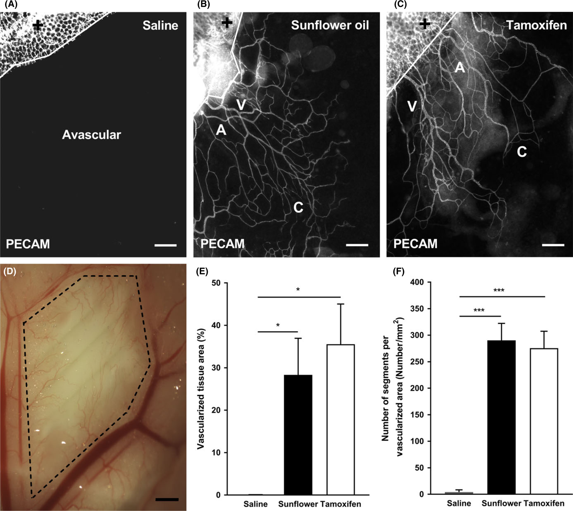
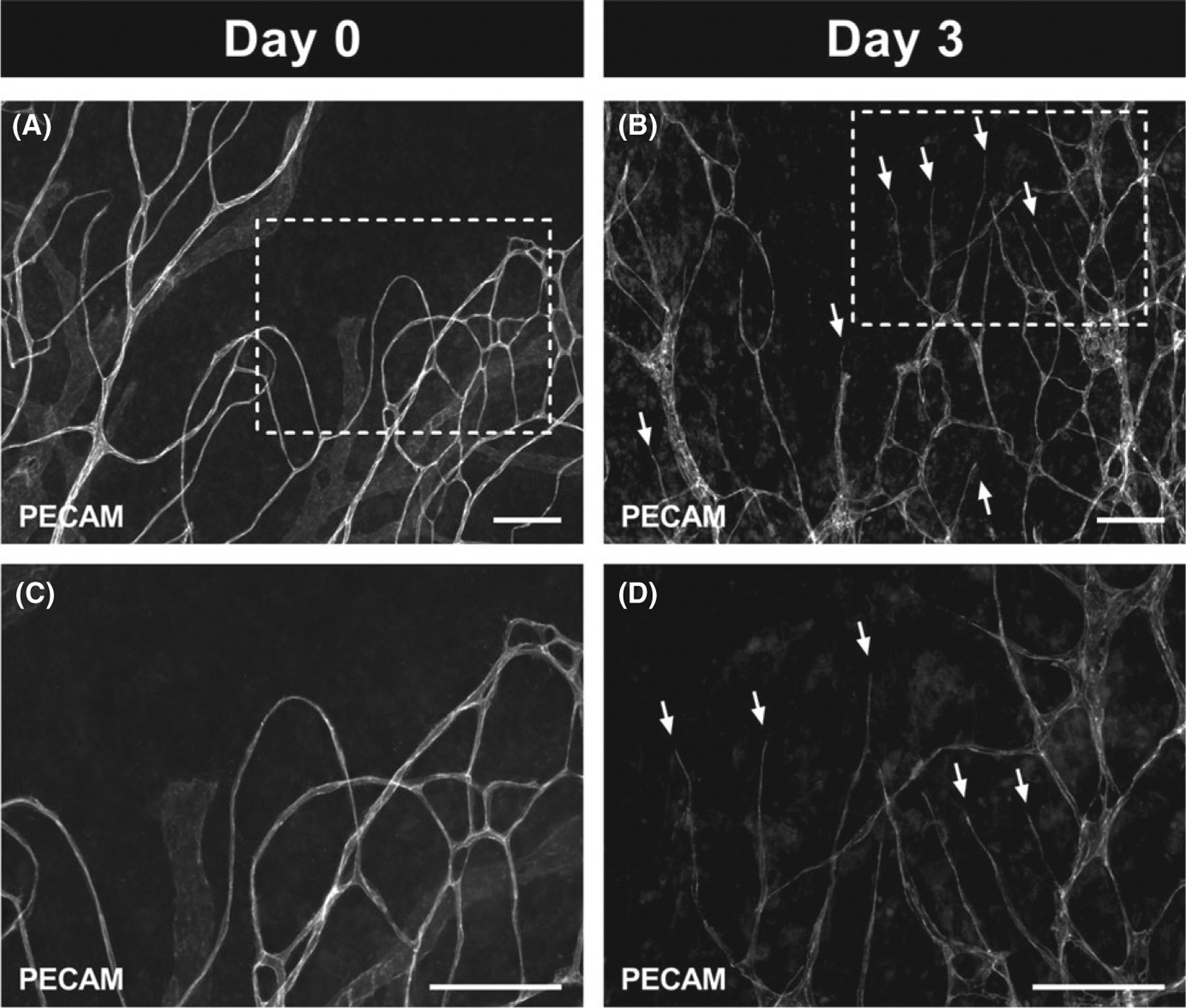
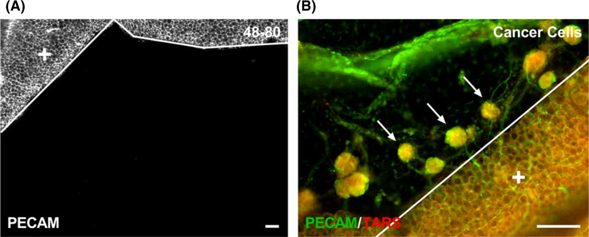
Objective: Motivated by observations of mesenteries harvested from mice treated with tamoxifen dissolved in oil for inducible gene mutation studies, the objective of this study was to demonstrate that microvascular growth can be induced in the avascular mouse mesentery tissue.

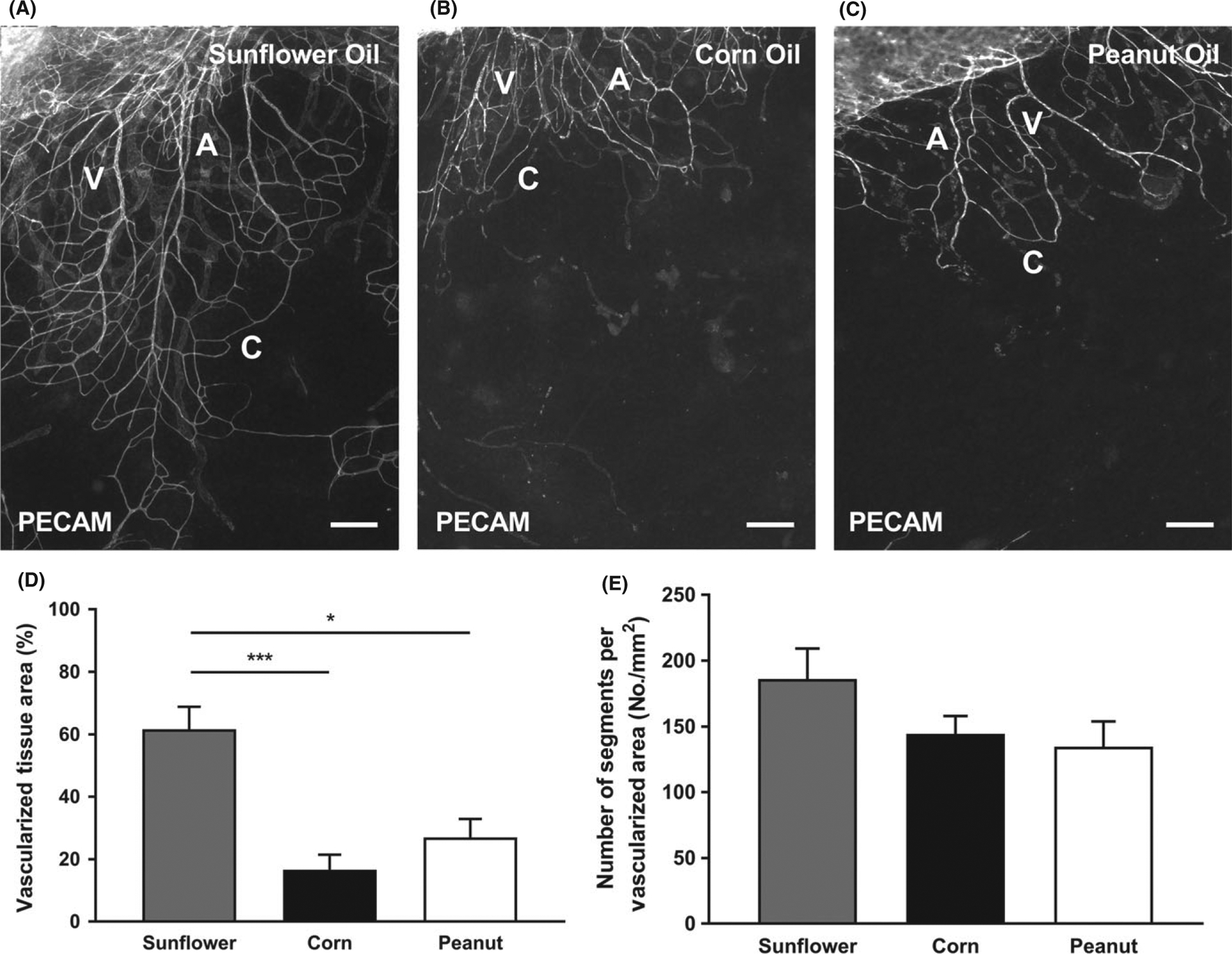
Methods: C57BL/6 mice were administered an IP injection for five consecutive days of: saline, sunflower oil, tamoxifen dissolved in sunflower oil, corn oil, or peanut oil.

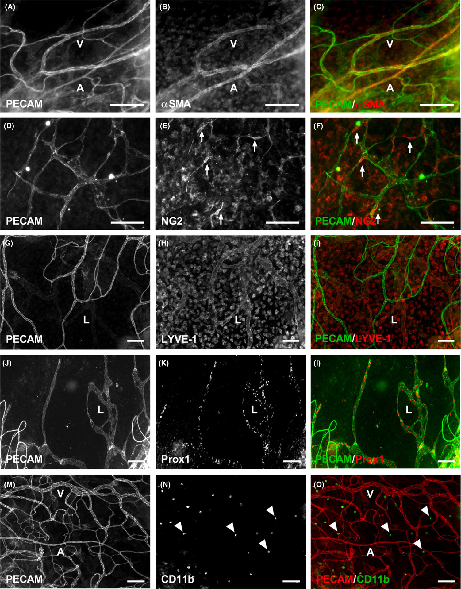
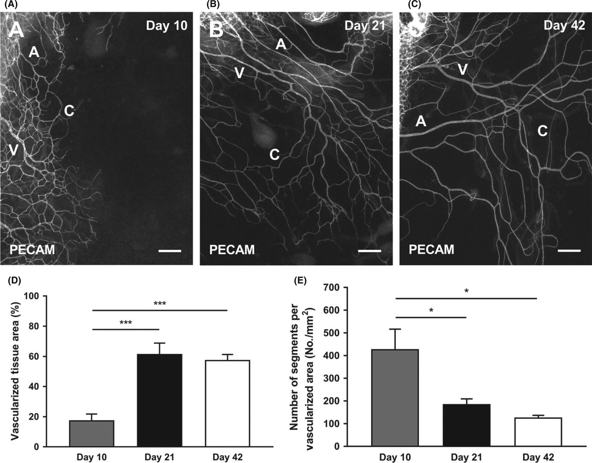
Results: Twenty-one days post-injection, zero tissues from saline group contained branching microvascular networks. In contrast, all tissues from the three oils and tamoxifen groups contained vascular networks with arterioles, venules, and capillaries. Smooth muscle cells and pericytes were present in their expected locations and wrapping morphologies. Significant increases in vascularized tissue area and vascular density were observed when compared to saline group, but sunflower oil and tamoxifen group were not significantly different. Vascularized tissues also contained LYVE-1-positive and Prox1-positive lymphatic networks, indicating that lymphangiogenesis was stimulated. When comparing the different oils, vascularized tissue area and vascular density of sunflower oil were significantly higher than corn and peanut oils.

Conclusions: These results provide novel evidence supporting that induction of microvascular network growth into the normally avascular mouse mesentery is possible.

Keywords: angiogenesis; lymphangiogenesis; microcirculation; microvascular network; mouse mesentery.


Conflict of interest statement

CONFLIC T OF INTEREST

None.

Figures

FIGURE 1
FIGURE 1
Growth of microvascular networks in mouse mesentery. PECAM labeling identified endothelial cells along microvascular networks. Mouse mesentery tissue from 21 d post-injection with saline (A), non-sterile sunflower oil (B), and tamoxifen (C); scale bars = 200 μm. The solid lines denote the border between the connective tissue and the adipose tissue (+). Bright field image shows perfused microvasculature (D) from sterile sunflower oil within the mesenteric window (dashed lines); scale bar = 1 mm. The quantification of vascularized tissue area (E) and vascular density (F) from each group is shown. A = arteriole, V = venule, and C = capillary. Data are shown as the mean + SEM, n = 8. The * and *** represent P < 0.05 and P < 0.0001, respectively
FIGURE 2
FIGURE 2
Multiple cell types are present in the vascularized mouse mesentery tissues 21 d post sunflower oil stimulation. Mouse mesentery tissues have PECAM-positive endothelial cells comprising microvascular networks (A-O), along with supporting cells including αSMA-positive smooth muscle cells (A-C), NG 2-positive pericytes (D-F), and CD 11b-positive macrophages (M-O). In addition to blood vessels, LYVE-1-positive (G-I) and Prox1-positive (J-L) lymphatic vessels are also present. A = arteriole, V = venule, and L = lymphatic. Arrows identify wrapping pericytes. Arrowheads identify interstitial macrophages. Scale bars = 100 μm
FIGURE 3
FIGURE 3
Growth of microvascular networks in mouse mesentery after 10, 21, and 42 d post sterile sunflower oil stimulation. PECAM labeling identified endothelial cells along microvascular networks Day 10 (A), Day 21 (B), and Day 42 (C) post-injection with sunflower oil. The quantification of vascularized tissue area (D) and vascular density (E) from the different days is shown. A = arteriole, V = venule, and C = capillary. Data are shown as the mean + SEM, n = 8. The * and *** represent P < 0.05 and P < 0.0001, respectively. Scale bars = 200 μm
FIGURE 4
FIGURE 4
Various sterile oil stimulations induce growth of microvasculature in the mouse mesentery. PECAM labeling identified endothelial cells along microvascular networks. Mouse mesentery tissue from 21 d post-injection with Sunflower Oil (A), Corn Oil (B), and Peanut Oil (C). The quantification of vascularized tissue area (D) and vascular density (E) from the different oils is shown. A = arteriole, V = venule, and C = capillary. Data are shown as the mean + SEM, n = 8. The * and *** represent P < 0.05 and P < 0.0001, respectively. Scale bars = 200 μm
FIGURE 5
FIGURE 5
Angiogenesis can be stimulated during culture in microvascular networks from mouse mesentery tissues after 21 d post sterile sunflower oil stimulation. Tissues cultured with MEM supplemented with 20% FBS for 3 d (B, D) had an obvious angiogenic response indicated by the capillary sprouts (arrows) when compared to tissues which had not been cultured (A, C). Images C and D are higher magnification of the images in A and B demarcated by the dashed rectangles. Scale bars = 100 μm
FIGURE 6
FIGURE 6
Different stimuli have different effects on microvascular growth in the mouse mesentery. After giving an IP injection of 0.5 mL doses of compound 48–80 (40, 80, 120, 160, and 200 μg/mL in sterile saline) over the time course of 3 d (twice a day and once on the last day) in increasing concentrations to adult, female C57 BL /6 mice (n = 2), PECAM-positive microvessels were not identified after 10 d post-injection (A). PECAM-positive endothelial cells comprising microvessels were observed after 4 wk of injected transformed mouse ovarian epithelial cells (3 × 10⁶ cells in 0.2 mL of PBS ), where spheroid tumors contained microvasculature into the mesenteric window (B). Arrows identify spheroid tumors which are comprised of TARS-positive cells. The solid lines denote the border between the connective tissue and the adipose tissue (+). Scale bars = 100 μm

References

    1. Breslin JW, Gaudreault N, Watson KD, Reynoso R, Yuan SY, Wu MH. Vascular endothelial growth factor-C stimulates the lymphatic pump by a VEGF receptor-3-dependent mechanism. Am J Physiol Heart Circ Physiol. 2007; 293:H709–H718. - PubMed
    1. Costa JJ, Harris AG, Delano FA, Zweifach BW, Schmid-schönbein GW. Mast cell degranulation and parenchymal cell injury in the rat mesentery. Microcirculation. 1999; 6:237–244. - PubMed
    1. Moazzam F, DeLano FA, Zweifach BW, Schmid-Schönbein GW. The leukocyte response to fluid stress. Proc Natl Acad Sci. 1997; 94 : 5338–5343. - PMC - PubMed
    1. Nehls V, Drenckhahn D. Heterogeneity of microvascular pericytes for smooth muscle type alpha-actin. J Cell Biol. 1991; 113:147–154. - PMC - PubMed
    1. Pearson MJ, Lipowsky HH. Influence of erythrocyte aggregation on leukocyte margination in postcapillary venules of rat mesentery. Am J Physiol Heart Circ Physiol. 2000; 279 : H1460–H1471. - PubMed

Publication types

LinkOut - more resources