Review
. 2018 Oct;126 Suppl 1(Suppl Suppl 1):37-48.
doi: 10.1111/eos.12536.

Current concepts on Sjögren's syndrome - classification criteria and biomarkers

Affiliations
Review

Current concepts on Sjögren's syndrome - classification criteria and biomarkers

Roland Jonsson et al. Eur J Oral Sci. 2018 Oct.

Abstract

Sjögren's syndrome is a lymphoproliferative disease with autoimmune features characterized by mononuclear cell infiltration of exocrine glands, notably the lacrimal and salivary glands. These lymphoid infiltrations lead to dryness of the eyes (keratoconjunctivitis sicca), dryness of the mouth (xerostomia), and, frequently, dryness of other surfaces connected to exocrine glands. Sjögren's syndrome is associated with the production of autoantibodies because B-cell activation is a consistent immunoregulatory abnormality. The spectrum of the disease extends from an organ-specific autoimmune disorder to a systemic process and is also associated with an increased risk of B-cell lymphoma. Current treatments are mainly symptomatic. As a result of the diverse presentation of the syndrome, a major challenge remains to improve diagnosis and therapy. For this purpose an international set of classification criteria for primary Sjögren's syndrome has recently been developed and validated and seems well suited for enrolment in clinical trials. Salivary gland biopsies have been examined and histopathology standards have been developed, to be used in clinical trials and patient stratification. Finally, ultrasonography and saliva meet the need of non-invasive imaging and sampling methods for discovery and validation of disease biomarkers in Sjögren's syndrome.

Keywords: Sjögren's syndrome; biomarkers; classification; histopathology; ultrasonography.


Conflict of interest statement

The authors declare that they have no conflict of interest.

Figures
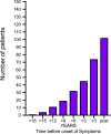
Figure 1
Figure 1
Number of patients, from a cohort of 171 patients, with positive anti‐nuclear antibody (ANA) score before (>18 yr) and after (post) onset of disease. Adapted from jonsson et al. 39 and theander et al. 40.
Figure 2
Figure 2
Ultrasonographic images of four left submandibular glands illustrating varying grades of normal/non‐specific to pathological changes. Grades 0–3 were used when evaluating ultrasonographic images of submandibular and parotid glands from each patient. (A) Grade 0. (B) Grade 1. (C) Grade 2. (D) Grade 3. Grades 0–1 were considered to correspond to normal morphology/non‐specific changes, whereas grades 2–3 were considered to correspond to pathological changes, possibly related to primary Sjögren's syndrome. Reproduced from hammenfors et al. 61, with permission.
Figure 3
Figure 3
Barriers and modes of transport for components originating from distant organs, the bloodstream, the interstitial space, acinar and ductal cells, or the oral cavity, to become detectable in saliva. The ability to monitor tissue‐related changes in saliva relies on the paradigm that a specific tissue state is reflected in the spectrum and quantity of specific components liberated into specific biofluids.
Figure 4
Figure 4
Schematic representation of the long covert phase of Sjögren's syndrome before disease onset followed by a commonly late diagnosis and currently limited options for biomarker‐based patient follow‐up. Ease of collection, repeatability, and close vicinity to the target organ make saliva a prime biofluid for biomarker discovery and clinical application in Sjögren's syndrome.
Figure 5
Figure 5
Salivary biomarker signatures serving in the diagnosis and stratification of patients as well as in predicting treatment responses in experimental models of Sjögrens syndrome. (A) 6‐plex [C‐reactive protein, interleukin (IL)‐4, pregnancy‐associated plasma protein A (PAPPA), IL‐5, clusterin, and apolipoprotein A2] salivary biomarker signature‐based recapitulation of the American–European Consensus Group criteria‐based classification. Discrimination between patients with Sjögren's syndrome, patients with rheumatoid arthritis, and asymptomatic controls was, at 94%, found to be highly accurate. Borrowed with permission from delaleu et al. 94. (B) 3‐plex [PAPPA, thrombospondin 1 (THBS‐1), and peptide YY (PYY)] biomarker signature‐based prediction of the nature of salivary gland inflammation for individual patients with Sjögren's syndrome. Borrowed with permission from delaleu et al. 95. A2M, alpha‐2‐macroglobulin; CCL2, chemokine (C‐C motif) ligand 2; CSF1, colony stimulating factor 1; CXCL9, chemokine (C‐X‐C motif) ligand 9; FS, focus score; GC, ectopic germinal centre; IL5, interleukin 5; INS, insulin; TT, transferrin; SELE, selectin E; SERPINE1, serpin family E member 1; SPP1, secreted phosphoprotein 1; UMOD, uromodulin. (C) Potential of a biomarker signature, combining granulocyte chemotactic protein 2 and IL‐1alpha measured in serum, and myeloperoxidase, myoglobin, and macrophage inflammatory protein 3b measured in saliva, to predict reinstatement of a physiological salivary secretion rate in a murine model of spontaneous Sjögren's syndrome after experimental treatment. Red and orange, responders to treatment; green, asymptomatic controls; purple and blue, non‐responders to treatment; black, disease control (accuracy = 93.8%). The x‐axis shows the salivary flow rate in microliters per minute per gram; the y‐axis represents the discriminant score of the biomarker signature. Borrowed with permission from delaleu et al. 96. aa, amino acids; deSF, decreased salivary flow; Hsp60, 60‐kd heat‐shock protein; IFA, Freund's incomplete adjuvant; NOD, nonobese diabetic mice; reSF, retained salivary flow. Circles represent individual patients (A, B) and mice (C).

References

    1. Sjögren H. Zur Kenntnis der Keratoconjunctivitis sicca. Acta Ophthalmol 1933; 11(Suppl): 1–151. - PubMed
    1. Chisholm DM, Mason DK. Labial salivary gland biopy in Sjögren's disease. J Clin Pathol 1968; 21: 656–660. - PMC - PubMed
    1. Greenspan JS, Daniels TE, Talal N, Sylvester RA. The histopathology of Sjögrens's syndrome in labial salivary gland biopsies. Oral Surg Oral Med Oral Pathol 1974; 37: 221–229. - PubMed
    1. Fisher BA, Jonsson R, Daniels T, Bombardieri M, Brown RM, Morgan P, Bombardieri S, Ng W‐F, Tzioufas AG, Vitali C, Shirlaw P, Haacke E, Costa S, Bootsma H, Devauchelle‐Pensec V, Radstake TR, Mariette X, Richards A, Stack R, Bowman SJ, Barone F. Standardisation of labial salivary gland histopathology in clinical trials in primary Sjögren's syndrome. Ann Rheum Dis 2017; 76: 1161–1168. - PMC - PubMed
    1. Shiboski CH, Shiboski SC, Seror R, Criswell LA, Labetoulle M, Lietman TM, Rasmussen A, Scofield H, Vitali C, Bowman SJ, Mariette X; International Sjögren's Syndrome Criteria Working Group . 2016 American College of Rheumatology/European League Against Rheumatism classification criteria for primary Sjögren's syndrome: A consensus and data‐driven methodology involving three international patient cohorts. Ann Rheum Dis 2017; 76: 9–16. - PubMed

Publication types