. 2018 Oct;592(19):3274-3285.
doi: 10.1002/1873-3468.13236. Epub 2018 Sep 20.

Extracellular histones are the ligands for the uptake of exosomes and hydroxyapatite-nanoparticles by tumor cells via syndecan-4

Affiliations

Extracellular histones are the ligands for the uptake of exosomes and hydroxyapatite-nanoparticles by tumor cells via syndecan-4

Josiah Ochieng et al. FEBS Lett. 2018 Oct.

Abstract

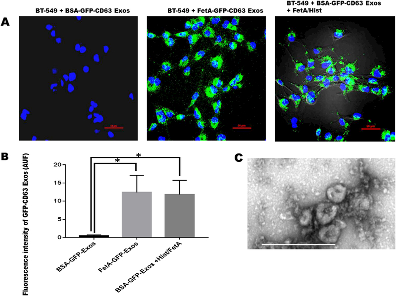
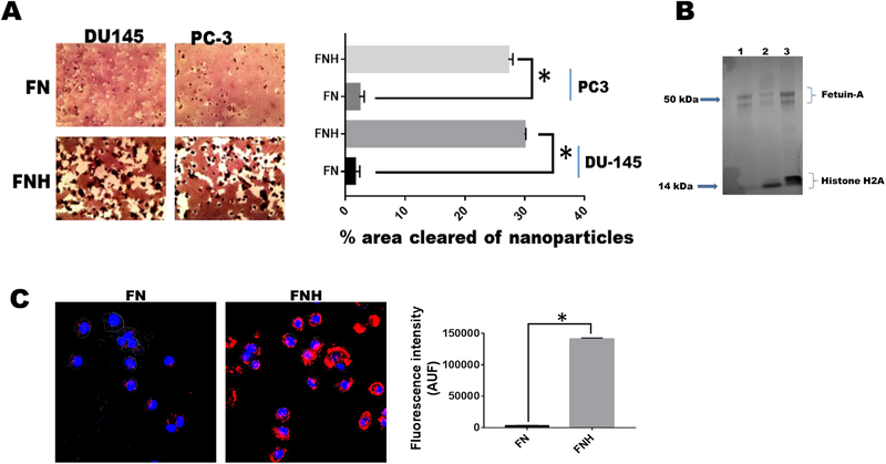
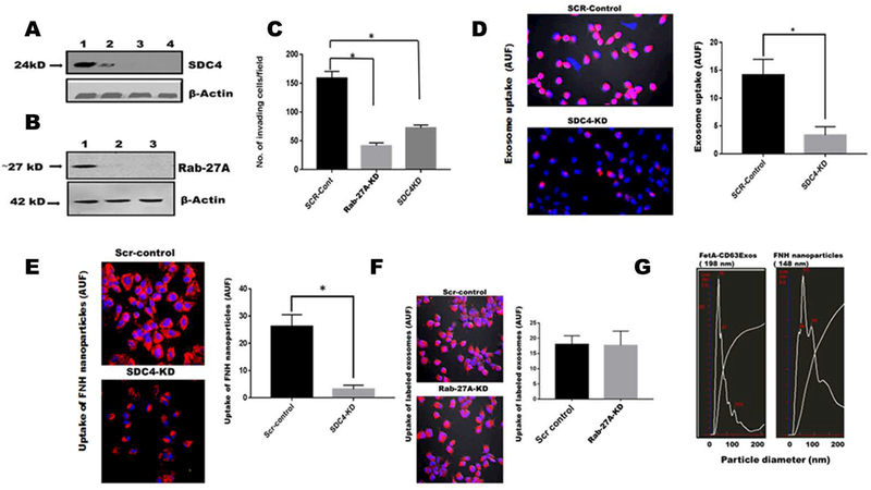
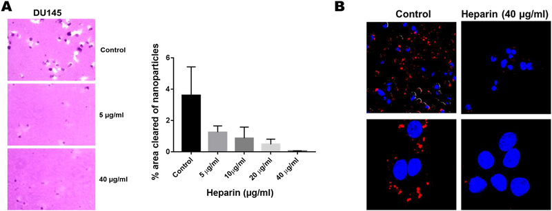
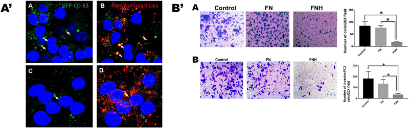
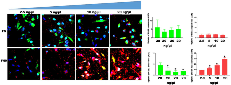
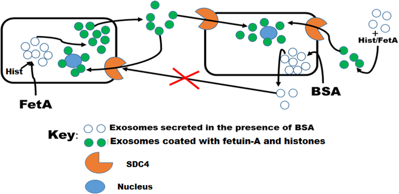
The mechanisms by which exosomes (nano-vesicular messengers of cells) are taken up by recipient cells are poorly understood. We hypothesized that histones associated with these nanoparticles are the ligands which facilitate their interaction with cell surface syndecan-4 (SDC4) to mediate their uptake. We show that the incubation with fetuin-A (exosome-associated proteins) and histones mediates the uptake of exosomes that are normally not endocytosed. Similarly, hydroxyapatite-nanoparticles incubated with fetuin-A and histones (FNH) are internalized by tumor cells, while nanoparticles incubated with fetuin-A alone (FN) are not. The uptake of exosomes and FNH, both of which move to the perinuclear region of the cell, is attenuated in SDC4-knockdown cells. Data show that FNH can compete with exosomes for uptake and that both use SDC4 as uptake receptors.

Keywords: exosomes; histones; hydroxyapatite; nanoparticles and syndecan-4.


Figures

Figure 1.
Figure 1.. Uptake of GFP-CD63 labeled exosomes by BT-549 breast carcinoma cells.
BT-539 cells were seeded in 8-chambered glass slides (1 × 10⁵ cells/chamber), and incubated for 24 h in complete medium and which the medium was replaced with SFM. Purified exosomes (100 μg/ml) secreted from BT-CD63 cells in the presence of BSA (negative control) or fetuin-A (positive control) in SFM were incubated with the BT-549 cells (2 μg/chamber) to monitor uptake. Exosomes secreted from BT-CD63 in the presence of BSA were also incubated with fetuin-A/histone H2A and re-purified (100 μg/ml) and finally incubated with BT-549 cells (2 μg/chamber) for uptake studies (Fig. 1A; scale bar = 50 μm). Mean arbitrary units of fluorescence ± SD were quantified by NEARS for each of the three measurements (Fig. 1B *P < 0.05; N = 6). Transmission electron micrograph of the purified exosomes obtained as described (14) is represented by Fig. 1C (scale bar is 500 nm).
Figure 2.
Figure 2.. Histones mediate the uptake of hydroxyapatite nanoparticles by tumor cells.
Hydroxyapatite-nanoparticles (2 mg/ml) were incubated with either 2 mg/ml of fetuin-A (FN) or fetuin-A (2 mg/ml) and histones (100 μg/ml) (FNH) in 10 ml of HBSS for 24 h at 4°C. The particles were centrifuged at 700 x g for 5 min to pellet large aggregated particles. The nanoparticles (colloidal) that remained suspended (9 ml) were washed 3X with HBSS, each time pelleted by centrifugation (5,000 x g for 5 min). The final pellets were suspended in 9 ml of complete medium and added to 96-well microtiter plates (100 μl/well) and the tumor cells (1,000 cells/well) added to the lawn of nanoparticles. After 48 h of incubation (37°C in humidified CO2 incubator), the cells were photographed and the % of areas cleared of nanoparticles quantified using NEARS (Panel A; *P < 0.001; N = 6). In panel B, the nanoparticles after the final wash in HBSS, were boiled in Laemmli sample buffer and resolved in NUPAGE and stained with colloidal Coomassie blue (lane 1-FN; lanes 2 and 3-FNH). In panel C, the fetuin-A was labeled with rhodamine isothiocyanate prior to incubation with hydroxyapatite nanoparticles (FN) or the nanoparticles and histones (FNH). The uptake of the nanoparticles (AUF) monitored using A1R confocal microscope and quantified by NEARS (*P < 0.001; N = 6).
Figure 3.
Figure 3.. Invasion and uptake of labeled exosomes and FNH nanoparticles by LN229 are attenuated in SDC4-knocked down cells.
Panel A, lane 1 (SCR-control); lane 2 (SDC4-KD-clone 2; lane 3 (SDC4-KD-clone 3 and lane 4 (SDC4-KD-clone 4). Panel B, lane 1 (SCR-control); lane 2 (Rab-27A-KD-clone 2); lane 3 (Rab-27A-KD-clone 3). In panel C, the invasion potentials of control cells transfected with scrambled shRNA (SCR-control) as well as clone 3 of Rab-27A-KD and SDC4-KD were assayed using Boyden chambers (Mean ± SD *P < 0.05; N = 7) In panel D, exosomes isolated and purified from BT-549 cells were labeled with rhodamine isothiocyanate and uptake of both SCR-control and SDC4-KD clone 3 cells determined (Nikon A1R) and AUF quantified by NEARS (Mean + SD, *P < 0.05; N = 6). Panel E, FNH nanoparticles were similarly labeled with rhodamine and uptake by SCR-controls and SDC4-KD cells determined as described for panel D. Panel F, uptake of rhodamine labeled FNH nanoparticles by SCR-controls and Rab-27A-KD cells was determined as described for panel D. Panel G, represents the particle size distribution of BT-CD63 exosomes and FNH nanoparticles as determined by NanoSight™.
Figure 4.
Figure 4.. Heparin inhibits the receptor mediated uptake of FNH nanoparticles.
In panel A, the areas cleared of nanoparticles in the absence or presence of heparin were quantified by NEARS. In panel B, the uptake of rhodamine labeled FNH nanoparticles by DU145 prostate cancer cells was determined by confocal microscopy (Nikon A1R). Lower panels represent zoomed images of the upper panels.
Figure 5.
Figure 5.. FNH nanoparticles and GFP-CD63 exosomes move to the perinuclear compartments of the cell and attenuate both motility and invasion of prostate cancer tumor cells.
In A’, GFP-CD63 exosomes (4 μg/chamber) and rhodamine labeled FNH nanoparticles (4 μg/chamber) were added to DU145 prostate cancer cells in 8-chambered glass slides. The green channel, panels A and C represent uptake of GFP-CD63 exosomes while the merged panels B and D show both GFP-CD63 exosomes and FNH nanoparticles which indicates that some of the exosomes were co-localized with FNH nanoparticles (arrows). In B’, the motility (panel A) and invasion (panel B) assays were done using Boyden Chambers as described. Briefly, the cells (DU145) were added to the upper chambers in SFM without (control) or with nanoparticles FN or FNH (100 μg/chamber). The lower chambers contained 500 μl of complete medium (10% fetal bovine serum). * P < 0.05, N = 6; one way ANOVA.
Figure 6.
Figure 6.. FNH nanoparticles competitively inhibits the uptake of exosomes by breast carcinoma cells.
MDA-MB-231 breast carcinoma cells were plated (2 × 10⁵ cells/chamber) in triplicates in 8-chambered glass slides. After an overnight attachment, complete medium in the chambers was replaced with SFM. GFP-CD63 labeled exosomes were added to each chamber (20 ng/μl) of three separate slides containing cells. To the upper four chambers of each slide, increasing concentrations of FN nanoparticles (2.5–20 ng/μl) in SFM were added. To the lower four chambers, increasing concentrations of FNH nanoparticles (2.5–20 ng/μl) in SFM were added. After 1 h of incubation at 37°C, the chambers were removed and the cells fixed in 4% formalin and a drop of slow-fade with Dapi added and cover-slipped. Uptake of exosomes (green channel) and nanoparticles (red channel) by the cells was monitored by confocal microscopy (Nikon A1R) and arbitrary units of fluorescence quantified using NEARS software as described in Materials and Methods. The bars represent means ± SD (*P < 0.05; N = 3; one way ANOVA)
Figure 7.
Figure 7.. Model depicting the uptake of exosome secreted in the absence or presence of fetuin-A.
In the presence of fetuin-A, the exosomes secreted are easily taken up by other tumor cells or by the secreting cells (autocrine). Exosomes secreted in the absence of fetuin-A but presence of BSA, lack the capacity to enter the cells via syndecan 4 (SDC4) receptors. Uptake incompetent exosomes secreted in the absence of fetuin-A can be incubated with a mixture of fetuin-A and histones and after further purification are now competent to enter the cells via SDC4.

Similar articles

Cited by

References

    1. Syn N, Wang L, Sethi G, Thiery JP & Goh BC (2016) Exosome-Mediated Metastasis: From Epithelial-Mesenchymal Transition to Escape from Immunosurveillance, Trends in pharmacological sciences. 37, 606–617. - PubMed
    1. Raposo G & Stoorvogel W (2013) Extracellular vesicles: exosomes, microvesicles, and friends, The Journal of cell biology. 200, 373–83. - PMC - PubMed
    1. Khushman M, Bhardwaj A, Patel GK, Laurini JA, Roveda K, Tan MC, Patton MC, Singh S, Taylor W & Singh AP (2017) Exosomal Markers (CD63 and CD9) Expression Pattern Using Immunohistochemistry in Resected Malignant and Nonmalignant Pancreatic Specimens, Pancreas. 46, 782–788. - PMC - PubMed
    1. Muhsin-Sharafaldine MR, Saunderson SC, Dunn AC, Faed JM, Kleffmann T & McLellan AD (2016) Procoagulant and immunogenic properties of melanoma exosomes, microvesicles and apoptotic vesicles, Oncotarget. 7, 56279–56294. - PMC - PubMed
    1. Skog J, Wurdinger T, van Rijn S, Meijer DH, Gainche L, Sena-Esteves M, Curry WT Jr., Carter BS, Krichevsky AM & Breakefield XO (2008) Glioblastoma microvesicles transport RNA and proteins that promote tumour growth and provide diagnostic biomarkers, Nature cell biology. 10, 1470–6. - PMC - PubMed

Publication types

MeSH terms