. 2019 Mar;69(3):1105-1121.
doi: 10.1002/hep.30249. Epub 2019 Feb 12.

Pharmacological Inhibition of CCR2/5 Signaling Prevents and Reverses Alcohol-Induced Liver Damage, Steatosis, and Inflammation in Mice

Affiliations

Pharmacological Inhibition of CCR2/5 Signaling Prevents and Reverses Alcohol-Induced Liver Damage, Steatosis, and Inflammation in Mice

Aditya Ambade et al. Hepatology. 2019 Mar.

Abstract

Kupffer cell and macrophage (MØ) activation contributes to steatosis, inflammation, and fibrosis in alcoholic liver disease (ALD). We found increased frequency of MØ, T cells, and expression of C-C chemokine receptor type 2 (Ccr2) and C-C chemokine receptor type 5 (Ccr5) in the livers of patients with ALD, and increased circulating chemokines, C-C chemokine ligand types 2 (CCL2), and C-C chemokine ligand types 5 (CCL5) in patients with alcoholic hepatitis. We hypothesized that inhibition of CCL2 signaling with the dual CCR2/5 inhibitor, cenicriviroc (CVC), would attenuate ALD. In a mouse model of ALD, liver injury (alanine aminotransferase [ALT]) and steatosis were prevented by CVC whether administered as "prevention" throughout the alcohol feeding or as "treatment" started after the development of ALD. Alcohol-induced increases in early liver fibrosis markers (sirius red, hydroxyproline, and collagen-1) were normalized by both modes of CVC administration. We found that prevention and treatment with CVC reversed alcohol-related increases in liver mRNA and protein expression of tumor necrosis factor (TNF)-α, interleukin (IL)-1β, IL-6, and CCL2. CVC administration regimens prevented the increase in infiltrating MØ (F4/80lo CD11bhi ) and reduced proinflammatory Ly6Chi MØ in livers of alcohol-fed mice. CVC increased liver T-cell numbers and attenuated Il-2 expression without an effect on CD69+ or CD25+ T-cell expression. In vitro, CVC inhibited CCL2-induced increases in hepatocyte fatty acid synthase (Fasn) and adipose differentiation-related protein (Adrp), whereas it augmented acyl-coenzyme A oxidase 1 (Acox-1), proliferator-activated receptor gamma co-activator alpha (Pgc1α) and uncoupling protein 2 expression, suggesting mechanisms for attenuated hepatocyte steatosis. We found that CCL2 and CCL5 sensitized hepatocytes to lipopolysaccharide-induced liver injury (TNF-α, ALT, and lactate dehydrogenase release). Alcohol feeding induced apoptosis (poly ADP-ribose polymerase [PARP] and caspase-3 [CASP-3] cleavage) and pyroptosis (gasdermin D [GSDMD] cleavage) in livers, and CVC prevented both of these forms of cell death. Conclusion: Together, our data demonstrate preclinical evidence for CCR2/CCR5 inhibition with CVC as a potent intervention to ameliorate alcohol-induced steatohepatitis and liver damage.


Figures

Figure 1.
Figure 1.. CCR2 and CCR5 expression and immune cell infiltration to the liver is increased in human alcoholic cirrhosis patients.
A) Chemokines CCL2, CCL3, CCL4 and CCL5 were measured from the serum of patients with alcoholic hepatitis or from healthy controls. B-C) Expression of chemokines Ccl2, Ccl3, Ccl4, Ccl5 Ccl8 and Ccl13 as well as chemokine receptors Ccr2 and Ccr5 were measured by qPCR from peripheral blood mononuclear cells (PBMCs) from patients with alcoholic hepatitis (B) and in liver tissue from patients with alcoholic cirrhosis (C) and compared with healthy controls. D) mRNA expression of macrophage markers Cd11b, Cd68 and Cd14 and T cell marker Cd3 was measured by qPCR in the livers of controls and patients with alcoholic cirrhosis. E) Liver tissue from controls and patients with alcoholic cirrhosis was immunohistochemically stained for the macrophage markers CD11b, CD68 and CD14 and the T cell marker CD3. * p<0.05 by student’s t-test
Figure 2.
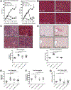
Figure 2.. Inhibition of CCR2/5 signaling alleviates chronic alcohol-induced liver injury.
Mice were fed a liquid diet containing 5% alcohol or pair-fed (PF) diet for 6 weeks. Some mice were injected subcutaneously with the CCR2/5 inhibitor Cenicriviroc (CVC) daily for the entire 6 weeks (6 wks) or during the final 3 weeks (wks 4–6) of ethanol feeding. Control mice were injected with vehicle. A) Serum ALT was measured weekly to track liver damage caused by chronic alcohol consumption. The Prevention Cohort received CVC daily beginning at the start of ethanol feeding while the Treatment Cohort began receiving injections at week 4. B) Livers from pair-fed (PF), alcohol-fed or alcohol-fed plus CVC treated mice were stained with hematoxylin and eosin. C) Oil Red O staining was completed on mouse livers to visualize and quantify lipid accumulation. D) Sirius Red staining was completed to visualize and quantify fibrosis development. E) Liver triglycerides were also measured. F) Liver hydroxyproline, a marker of collagen content, was measured using a colorimetric assay. G) Liver expression of collagen mRNA was measured by qPCR. * p<0.05 by student’s t-test in (A) and one-way ANOVA in (B-G).
Figure 3.
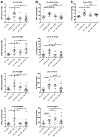
Figure 3.. CVC inhibits pro-inflammatory cytokine expression induced by alcohol.
A-B) Liver mRNA expression of pro-inflammatory cytokines Tnfa, Il-1β, Il-6 and Ccl2 were measured by qPCR (A) and liver protein was measured by ELISA (B). C) Serum soluble CD14 (sCD14) was measured by ELISA from mice fed alcohol, alcohol plus CVC and pair-fed mice. * p<0.05 by one-way ANOVA.
Figure 4.
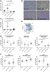
Figure 4.. CVC decreases the infiltration of peripheral macrophages into the liver.
A) Liver mRNA expression of Ccr2, a target of CVC, was measured by qPCR. B-C) Liver sections were formalin fixed and stained by immunohistochemistry for macrophage surface marker F4/80 to identify and quantify hepatic macrophages. D) Liver mononuclear cells were isolated via tissue digestion followed by density gradient, surface staining for CD11b and F4/80 for differentiating liver macropahges by flow cytometry. Cells were gated based on size, singlets, an amino-reactive dye for living cells and for CD45+ positivity. E) Flow cytometry was used to quantify total liver macrophages (F4/80+CD11b+), resident Kupffer cells (F4/80hiCD11blo) as well as infiltrating liver macrophages (F4/80loCD11bhi). F) Infiltrating F4/80loCD11bhi macrophages were further differentiated by Ly6CLo, Ly6CMed and Ly6CHi expression from pair-fed, alcohol-fed and alcohol-fed CVC “treatment” mice. HPF: high-power field. * p<0.05 by one-way ANOVA.
Figure 5.
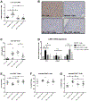
Figure 5.. CVC reduces the infiltration and activation of T cells into the liver following chronic alcohol.
A) Liver mRNA expression of Ccr5, another target of CVC, was measured by qPCR. B-C) Liver sections were formalin fixed and stained by immunohistochemistry for T cell surface receptor CD3 to identify and quantify hepatic T cells. D) Liver mononuclear cells were isolated via tissue digestion followed by density gradient and mRNA expression of T cell-associated genes Il-2, Foxp3 and Ifnγ were measured by qPCR. E-G) Flow cytometry was used to quantify total liver T cells (E; CD3+), activated CD69+ T cells (F) as well as CD25+ T cells (G). Cells were gated for lymphocytes, singlets, an amino-reactive dye for living cells and for CD45+/CD3+ positivity. * p<0.05 by one-way ANOVA.
Figure 6.
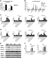
Figure 6.. CVC protects from induction of steatosis, inflammation and cell death in primary hepatocytes.
A) Expression of receptor proteins CCR2 and CCR5 was measured on primary mouse hepatocytes from animals fed alcohol or a calorie-matched diet. Both were elevated by chronic alcohol consumption. B) To assess the influence of signaling through CCR2, primary hepatocytes were isolated and cultured in vitro with either recombinant CCL2 alone, CVC alone or CCL2 with CVC. mRNA expression of genes associated with fatty acid metabolism revealed the effects of CCL2 and CVC. C-D) Either CCL2 (C) or CCL5 (D), ligands of CCR2 and CCR5 respectively, were used in combination with varying doses of LPS and CVC to observe the induction of TNFα as well as indicators of cell death including alanine aminotransferase (ALT), lactate dehydrogenase release and caspase-3 activity. CVC protected hepatocytes from damage in a dose-dependent manner. E) Western blot measurement of liver lysates from alcohol-fed mice without and with CVC administration as well as pair-fed mice was completed and probed for full (fl) and cleaved (clv) poly ADP-ribose polymerase (PARP), caspase-3, caspase-11 and gasdermin D (GSDMD). F) Primary mouse bone-marrow derived macrophages were treated in vitro with CVC (5nM, 20nM and 100nM) then LPS (10ng/mL) and proinflammatory cyotkines TNFα and IL-6 were measured from cell supernatants. * p<0.05 by student’s t-test or by one-way ANOVA.

References

    1. O’Shea RS, Dasarathy S, McCullough AJ. Alcoholic liver disease. Hepatology 2010;51:307–328. - PubMed
    1. Schwartz JM, Reinus JF. Prevalence and natural history of alcoholic liver disease. Clin Liver Dis 2012;16:659–666. - PubMed
    1. Lucey MR, Mathurin P, Morgan TR. Alcoholic hepatitis. N Engl J Med 2009;360:2758–2769. - PubMed
    1. Frazier TH, Stocker AM, Kershner NA, Marsano LS, McClain CJ. Treatment of alcoholic liver disease. Therap Adv Gastroenterol 2011;4:63–81. - PMC - PubMed
    1. Gao B, Bataller R. Alcoholic liver disease: pathogenesis and new therapeutic targets. Gastroenterology 2011;141:1572–1585. - PMC - PubMed

Publication types

MeSH terms