. 2018 Oct;84(4):576-587.
doi: 10.1002/ana.25324. Epub 2018 Oct 5.

Association between carotid atheroma and cerebral cortex structure at age 73 years

Affiliations

Association between carotid atheroma and cerebral cortex structure at age 73 years

Saud Alhusaini et al. Ann Neurol. 2018 Oct.

Abstract

Objective: To examine the relationship between carotid atherosclerosis and cerebral cortical thickness and investigate whether cortical thickness mediates the association between carotid atheroma and relative cognitive decline.

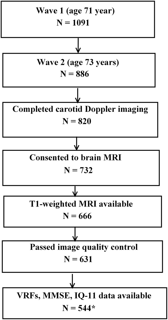
Methods: We assessed 554 community-dwelling subjects (male/female: 296/258) from the Lothian Birth Cohort 1936 who underwent brain magnetic resonance imaging and carotid Doppler ultrasound studies at age 73 years. The relationship between carotid atherosclerosis markers (internal carotid artery stenosis, intima-media thickness, velocity, pulsatility, and resistivity indexes) and vertex-wide cerebral cortical thickness was examined cross-sectionally, controlling for gender, extensive vascular risk factors (VRFs), and intelligence quotient at age 11 (IQ-11). We also determined the association between carotid stenosis and a composite measure of fluid intelligence at age 73 years. A mediation model was applied to examine whether cortical thickness mediated the relationship between carotid stenosis and cognitive function.

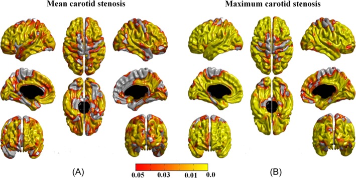
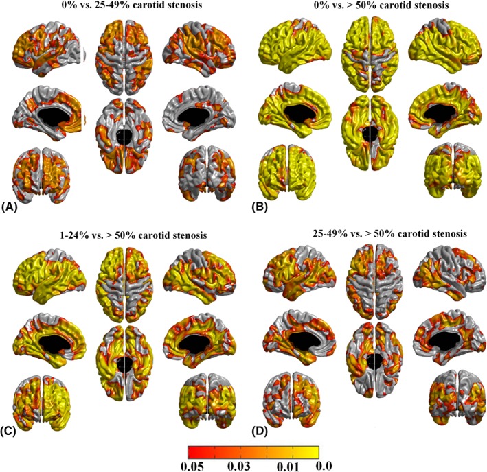
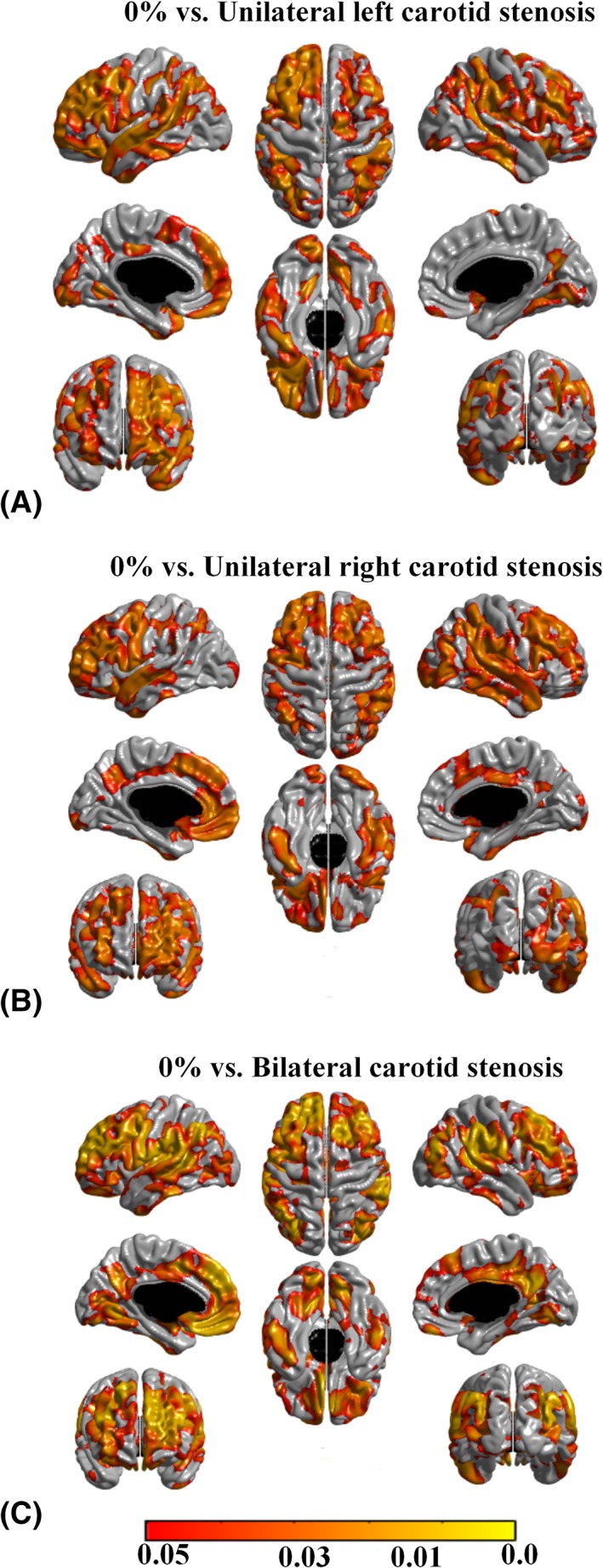
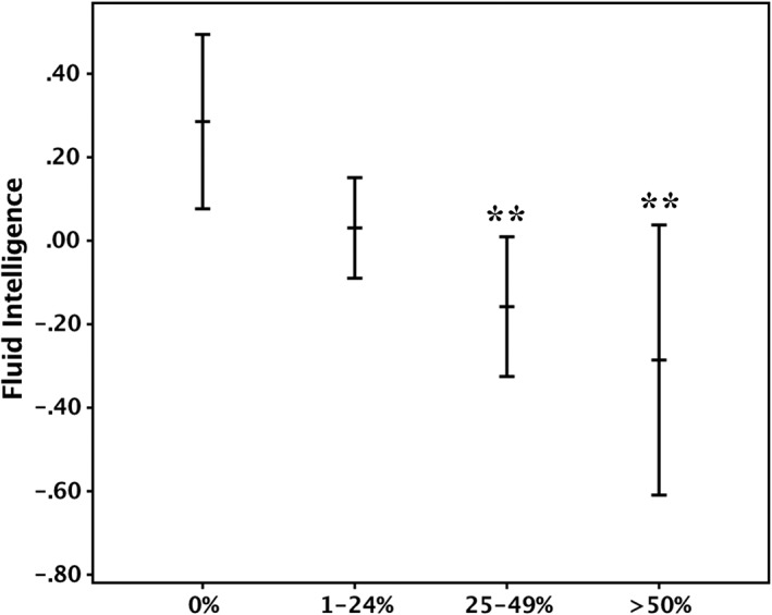
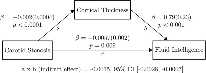
Results: A widespread negative association was identified between carotid stenosis (median = 15%) and cerebral cortical thickness at age 73 years, independent of the side of carotid stenosis, other carotid measures, VRFs, and IQ-11. This association increased in an almost dose-response relationship from mild to severe degrees of carotid stenosis, across the anterior and posterior circulation territories. A negative association was also noted between carotid stenosis and fluid intelligence (standardized beta coefficient = -0.151, p = 0.001), which appeared partly (approximately 22%) mediated by carotid stenosis-related thinning of the cerebral cortex.

Interpretation: The findings suggest that carotid stenosis represents a marker of processes that accelerate aging of the cerebral cortex and cognition that is in part independent of measurable VRFs. Cortical thinning within the anterior and posterior circulation territories partially mediated the relationship between carotid atheroma and fluid intelligence. Ann Neurol 2018;84:576-587.


Figures

Figure 1
Figure 1
Flowchart indicating the selection process of the Lothian Birth Cohort 1936 subjects included in the final sample for data analyses. *Final sample included in the cross‐sectional analyses reported here. IQ‐11 = intelligence quotient at 11 years; MMSE = Mini‐Mental State Examination; MRI = magnetic resonance imaging; VRF = vascular risk factor.
Figure 2
Figure 2
The associations between carotid stenosis and cortical thickness. (A) The association between mean carotid stenosis (average of the left and right internal carotid artery [ICA] stenosis) and cortical thickness, controlling for age (in days) and gender. (B) The association between maximum carotid stenosis (observed on the left or right ICA) and cortical thickness, controlling for age (in days) and gender. The associations in A and B remained significant after controlling for vascular risk factors, intelligence quotient at age 11 years, Mini‐Mental State Examination score, and other carotid measures, affecting both carotid territory and regions supplied by the posterior circulation that are anatomically unrelated to the carotid arteries. Areas in orange–yellow shades represent statistically significant negative associations at false discovery rate (FDR) = 0.05. The color bar represents FDR q values.
Figure 3
Figure 3
Pattern of cerebral cortical thinning in (A) individuals with 25–49% carotid stenosis compared to those with no carotid stenosis, (B) individuals with ≥ 50% carotid stenosis compared to those with no carotid stenosis, (C) individuals with ≥ 50% carotid stenosis compared to those with 1–24% carotid stenosis, and (D) individuals with ≥ 50% carotid stenosis compared to those with 25–49% carotid stenosis. Age (in days), gender, vascular risk factors, intelligence quotient at age 11 years, Mini‐Mental State Examination score, and other carotid measures were included as covariates. Areas in orange–yellow shades represent statistically significant cortical thinning at a false discovery rate (FDR) = 0.05. The color bar represents FDR q values. The group contrasts between 0% and 1–24% carotid stenosis and between 1–24% and 25–49% carotid stenosis did not reach statistical significance.
Figure 4
Figure 4
Pattern of cerebral cortical thickness reduction in (A) individuals with ≥ 25% left carotid stenosis compared to those with no carotid stenosis, (B) individuals with ≥ 25% right carotid stenosis compared to those with no carotid stenosis, and (C) individuals with ≥ 25% bilateral carotid stenosis compared to those with no carotid stenosis. Areas in orange–yellow shades represent statistically significant cortical thinning at a false discovery rate (FDR) = 0.05. The color bar represents FDR q values.
Figure 5
Figure 5
Mean fluid intelligence score in individuals with 0%, 1–24%, 25–49%, and ≥50% carotid stenosis. Error bands represent 95% confidence intervals. The score was derived using a principal component analysis from the following subsets of the Wechsler Adult Intelligence Scale, 3rd UK edition (WAIS‐III): Matrix Reasoning, Block Design, Digit Span Backward, Letter‐Number Sequencing, Digit Symbol Substitution, and Symbol Search. **p < 0.01 (comparisons were made against individuals with 0% carotid stenosis). Covariates: age, gender, vascular risk factors, and intelligence quotient at age 11 years.
Figure 6
Figure 6
The result of the mediation model applied to examine the effect of cortical thickness on the relationship between carotid stenosis and fluid intelligence is displayed. To investigate the association between carotid atherosclerosis, cortical thickness, and fluid intelligence, (1) maximum carotid stenosis was used as a marker for carotid atherosclerosis; (2) the average thickness of the significant cortical areas derived from the analysis of the relationship between cortical thickness and carotid stenosis was used for the cortical thickness variable (shown in Fig 2B); and (3) the principal component analysis–derived general component from the following subsets of the Wechsler Adult Intelligence Scale, 3rd edition was used for the fluid intelligence variable: Matrix Reasoning, Block Design, Digit Span Backward, Letter‐Number Sequencing, Digit Symbol Substitution, and Symbol Search. Each arrow is directed from a predictor variable to the outcome variable: (1) the relationship between carotid stenosis and cortical thickness is denoted by path a; (2) the relationship between cortical thickness and fluid intelligence, adjusted for carotid stenosis, is denoted by path b; (3) the direct effect of carotid stenosis upon fluid intelligence is denoted by path c′, and was measured after adjustment for cortical thickness, gender, intelligence quotient at age 11 years, and all vascular risk factors. Beta coefficients and related p values displayed adjacent to each path represent unstandardized regression coefficients (standard errors) generated (an ordinary least squares regression model) as part of the mediation model using the PROCESS macro for SPSS. A formal test of the significance of the mediation effect was carried out, for which the results are listed at the bottom of the figure. The indirect effect in the mediation model (the degree to which the effect of carotid stenosis on fluid intelligence is believed to be transmitted through mean cortical thickness) was assessed using a bias‐corrected bootstrapped confidence interval (CI) derived by Monte Carlo sampling. Number of bootstrap samples = 10,000.

Similar articles

Cited by

References

    1. Tsivgoulis G, Vemmos K, Papamichael C, et al. Common carotid artery intima‐media thickness and the risk of stroke recurrence. Stroke 2006;37:1913–1916. - PubMed
    1. Muller M, Grobbee DE, Aleman A, et al. Cardiovascular disease and cognitive performance in middle‐aged and elderly men. Atherosclerosis 2007;190:143–149. - PubMed
    1. Bots ML, Breslau PJ, Briet E, et al. Cardiovascular determinants of carotid artery disease. The Rotterdam Elderly Study. Hypertension 1992;19:717–720. - PubMed
    1. Wardlaw JM, Allerhand M, Doubal FN, et al. Vascular risk factors, large‐artery atheroma, and brain white matter hyperintensities. Neurology 2014;82:1331–1338. - PMC - PubMed
    1. Romero JR, Beiser A, Seshadri S, et al. Carotid artery atherosclerosis, MRI indices of brain ischemia, aging, and cognitive impairment: the Framingham study. Stroke 2009;40:1590–1596. - PMC - PubMed

Publication types