. 2018 Nov:183:280-294.
doi: 10.1016/j.biomaterials.2018.08.053. Epub 2018 Aug 30.

Tumor hypoxia directed multimodal nanotherapy for overcoming drug resistance in renal cell carcinoma and reprogramming macrophages

Affiliations

Tumor hypoxia directed multimodal nanotherapy for overcoming drug resistance in renal cell carcinoma and reprogramming macrophages

Hashem O Alsaab et al. Biomaterials. 2018 Nov.

Abstract

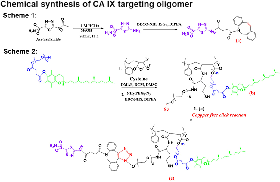
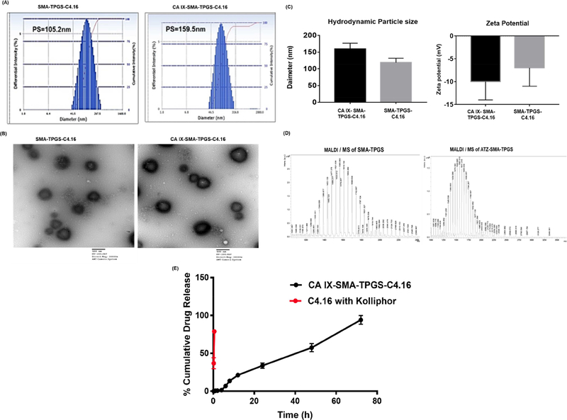
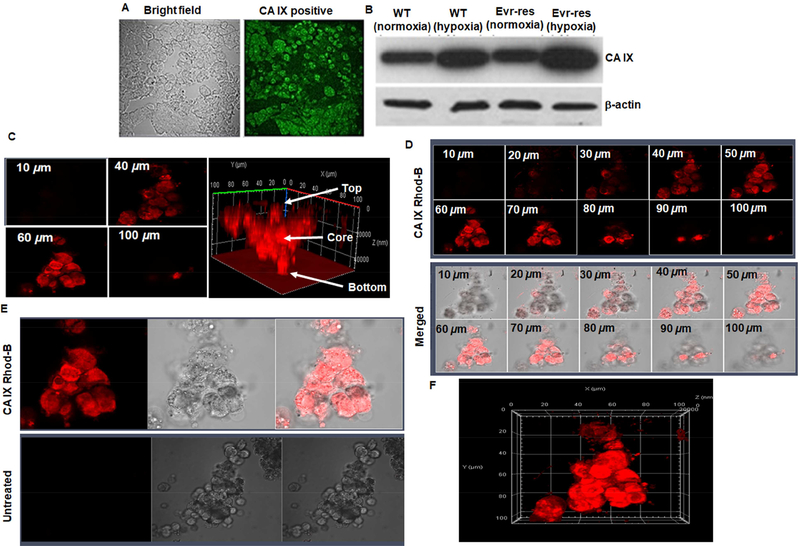
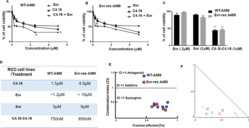
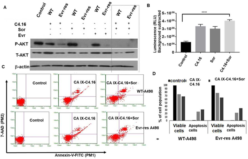
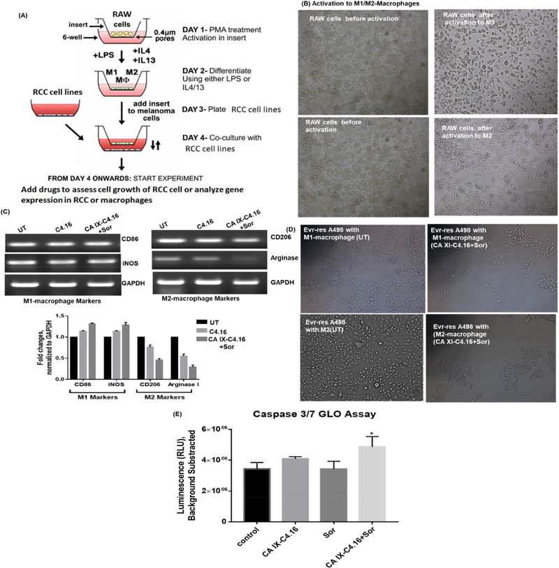
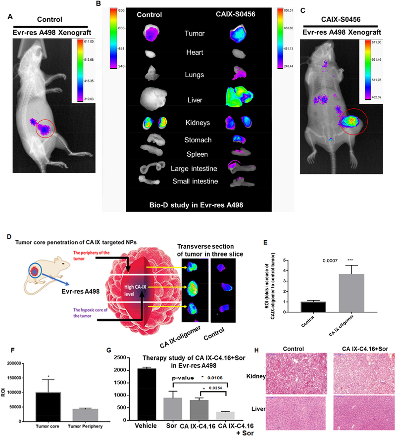
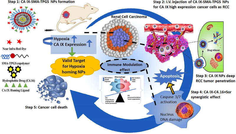
Drug resistance is one of the significant clinical burden in renal cell carcinoma (RCC). The development of drug resistance is attributed to many factors, including impairment of apoptosis, elevation of carbonic anhydrase IX (CA IX, a marker of tumor hypoxia), and infiltration of tumorigenic immune cells. To alleviate the drug resistance, we have used Sorafenib (Sor) in combination with tumor hypoxia directed nanoparticle (NP) loaded with a new class of apoptosis inducer, CFM 4.16 (C4.16), namely CA IX-C4.16. The NP is designed to selectively deliver the payload to the hypoxic tumor (core), provoke superior cell death in parental (WT) and Everolimus-resistant (Evr-res) RCC and selectively downmodulate tumorigenic M2-macrophage. Copper-free 'click' chemistry was utilized for conjugating SMA-TPGS with Acetazolamide (ATZ, a CA IX-specific targeting ligand). The NP was further tagged with a clinically approved NIR dye (S0456) for evaluating hypoxic tumor core penetration and organ distribution. Imaging of tumor spheroid treated with NIR dye-labeled CA IX-SMA-TPGS revealed remarkable tumor core penetration that was modulated by CA IX-mediated targeting in hypoxic-A498 RCC cells. The significant cell killing effect with synergistic combination index (CI) of CA IX-C4.16 and Sor treatment suggests efficient reversal of Evr-resistance in A498 cells. The CA IX directed nanoplatform in combination with Sor has shown multiple benefits in overcoming drug resistance through (i) inhibition of p-AKT, (ii) upregulation of tumoricidal M1 macrophages resulting in induction of caspase 3/7 mediated apoptosis of Evr-res A498 cells in macrophage-RCC co-culturing condition, (iii) significant in vitro and in vivo Evr-res A498 tumor growth inhibition as compared to individual therapy, and (iv) untraceable liver and kidney toxicity in mice. Near-infrared (NIR) imaging of CA IX-SMA-TPGS-S0456 in Evr-res A498 RCC model exhibited significant accumulation of CA IX-oligomer in tumor core with >3-fold higher tumor uptake as compared to control. In conclusion, this proof-of-concept study demonstrates versatile tumor hypoxia directed nanoplatform that can work in synergy with existing drugs for reversing drug-resistance in RCC accompanied with re-education of tumor-associated macrophages, that could be applied universally for several hypoxic tumors.

Keywords: Carbonic anhydrase IX; Everolimus; Macrophage modulation; Nano-therapy; Overcoming drug resistance; Renal cell carcinoma; Tumor core penetration; Tumor hypoxia targeting.


Conflict of interest statement

Declarations of interest

There are no conflicts to declare.

Figures

Figure 1:
Figure 1:
The general procedure for acetazolamide-oligomer (CA IX-SMA-TPGS) synthesis is shown. The final ‘click’ reaction product, compound c has been used to encapsulate C4.16 or to conjugate with S0456 or Rhodamine dye with -SH functional group to obtain CA IX-oligomer.
Figure 2.
Figure 2.. Nanoparticle formulation and characterization.
(A) Hydrodynamic size of targeted non-targeted SMA-TPGS-C4.16 and hypoxia targeting CA IX-SMA-TPGSC4.16 NP are shown. (B) The transmission electron micrographs shows the morphology of non-targeted and targeted NP. (C) The zeta potential or surface charge on the NPs by Dynamic Light Scattering (DLS) is shown. (Representative histogram of hydrodynamic particle size and zeta potential (n=3). (D) MALDI/MS analysis of CA IXSMA-TPGS and SMA-TPGS are shown. The increment of molecular weight in CA IXSMA-TPGS (m/z 3126) compared to SMA-TPGS (m/z 2399), and their corresponding fragmented peaks indicates the successful conjugation of ATZ to the SMA-TPGS polymer. (E) In vitro drug release kinetics of CAIX-SMA-TPGS-C4.16 in PBS indicates that the sustained release of C4.16 from the CAIX-SMA-TPGS-C4.16 NP as compared to free C4.16 with excipient, such as Kolliphor in PBS is shown.
Figure 3.
Figure 3.. Hypoxia induced CA IX-overexpression in A498 cells and tumors to enable tumor core penetration of CA IX oligomer.
(A) Immunohistochemistry of CA IX-positive A498 RCC tumor xenografts collected from tumor tissue section is shown. The intense bright green fluorescence indicates the rationale for choosing CA IX as an excellent biomarker for RCC specific payload delivery. (B) Western blot detection of CA IX protein in A498 and EV-A498 RCC cells lysates after normoxia and hypoxia (treated with cobalt chloride for 72 h) are shown. The fold up-regulation of CA IX expression in hypoxic WT and EV-res A498 RCC cells compared to normoxia provides a solid foundation for delivering the payload into oxygen-deprived regions and the hypoxic core of RCC tumor. (C) 3D spheroid uptake studies of hypoxia targeted-oligomer (CA IX Rhod-B). Confocal microscope images of CA IX Rhod-B treated hypoxic A498 spheroid indicates tumor matrix penetration of CA IX-oligomer. The untreated and treated spheres were then photographed as noted in the methods section. Z-stacking of the spheroid clearly indicates that fluorescence intensity is superior in 40–60 μm section (core) as compared to 10 or 100 μm (periphery). The highest fluorescence intensity at the center (as indicated by arrow) of 3D- plot suggests that CA IX-Rhod oligomer is highly efficient to reach deep into the core of the tumor spheroid. (D) Z-stacking of the spheroid at different sections from 10–100μm with CA IX targeted formulations also reveals similar findings as noted for the 40–60 μm that had superior fluorescence intensity. Figure (E) shows the untreated control experiments in comparison with CA IXRhod oligomer and Figure (F) shows the overall shape and morphology of the spheroid along the three dimensional (x, y, and z) axis.
Figure 4.
Figure 4.. C4.16 and CA IX-C4.16 are more efficient in inhibiting the growth of WT and Evr-res A498 cells.
In vitro cytotoxicity assay of C4.16 and Sor on (A) WT and (B) Evr-res A498 indicates C4.16 was more potent than the FDA approved drug, Sor and combining both drugs C4.16+Sor demonstrated significantly lower cell viability. (C) The results also showed that CA IX-C4.16 is more effective in inhibiting the growth of A498 (WT and Evr-res) RCC cell lines compared to Sor and Evr and support the notion that C4.16 is more potent than FDA approved drugs in the RCC model. (D) Summary of IC50 value for all the tested drugs with the tested RCC cell lines are shown in a tabular fashion. The data in the IC50 columns represent the mean of three independent experiments. Indicated A498 WT and their respective Evr-res A498 cells were either untreated (control) or treated with a noted dose of C4.16, Sor, Evr, and CA IX-C4.16 for 48 h. (E) High synergistic CI value of C4.16 in combination with Sor supports the hypothesis of selecting the combination to treat RCC for reversing the drug resistance. This data builds a rationale for using hypoxic core penetrating CA IX-C4.16+Sor to sensitize the drug resistant RCC. (F) Isobologram of CA IX-C4.16+Sor suggests high synergism combination treatment in RCC cells.
Figure 5.
Figure 5.. (A) Molecular mechanism of cell death and resurrection of apoptosis:
Western blot analysis clearly indicates that C4.16+Sor combination completely wiped out P-AKT level both in WT and Evr-res A498 cells. Cell were either kept untreated (control, C) or treated with Evr, Sor, C4.16 or C4.16+Sor. (B) Up-regulation of caspase 7/9 with (C4.16+Sor) treatment in Evr-res A498 cells indicates effective induction of apoptosis to drug resistant cells as compared to control or individual treatment. The results support the notion that (C4.16+Sor) combination is more effective in resurrecting apoptosis mediated cell death. Data represent mean ± SD, n=3 per group, ****p<0.01 vs. control. (C) Apoptosis analysis of WT and Evr-res A498 cell by FACS using dual Annexin-V and 7-AAD staining. The data indicates CA IX-C4.16+Sor is superior in inducing apoptosis as compared to control; CA IX-C4.16 NP treated cells. (D) Histogram columns of both viable cells and apoptotic cells indicates that CA IX-C4.16 + Sor has more % apoptotic cell compared to CA IX-C4.16 alone which support our hypothesis of the synergism in RCC cell killing.
Figure 6.
Figure 6.. Reprogramming macrophages with CA IX-C4.16+Sorafenib treatment.
(A) Schematic representation of the procol is shown. Raw-264.7 cells were placed into the insert. Then, cells were polarized to M1-macrophage using IFN-γ and LPS, and to M2-macrophage using IL-4 recombinant protein. The scheme was modified from the original protocol by Smith et al. (B) Change of morphology of M1 and M2 macrophages supports the polarization of Raw-264.7. (C) RT-PCR data clearly demonstrates the upmodulation of the tumoricidal M1-macrophage marker (CD86, iNOS) and downmodulation of the tumorigenic M2-macrophage marker (CD206, Arginase I) in CA IXC4.16+Sor as compared to control and C4.16. The macrophage reprogramming ability of CA IX-targeting NP builds a rational of using (CA IX-C-4.16+Sor) as a potent antitumor immune-stimulatory agent for RCC. (D) Change of morphology and reduction of Evr-res A498 density in M1-macrophage and Evr-res A498 co-cultured condition, treated with CA XI+Sor suggests activated M1-macrophage mediated RCC cell death. (E) Treatment of CA IX+Sor educate the Raw-264.7 in inducing caspase 3/7 mediated apoptosis of Evr-res A498.
Figure 7:
Figure 7:. Superior tumor specificity of CA IX-oligomer and combination antitumor efficacy study in Evr-res A498 xenograft RCC model.
(A and C) Superior tumor accumulation of CAIX oligomer (CA IX-S0456) as compared to control (S0456) in Evrres A498 tumor xenograft model is shown. (B) Biodistribution (Bio-D) study of CA IXS0456 showed superior tumor specificity and low non-specific liver uptake in Evr-res A498 tumor bearing mice. The control, S0456 showed poor tumor accumulation with high off-target activity. (D) Further to demonstrate the tumor core penetration of NIR dye, the isolated Evr-res A498 tumor was transversely sectioned into 3 parts; the brightest fluorescence intensity at the middle (core) section confirmed the excellent hypoxic tumor core penetration ability of CA IX-S0456 compared with non targeted control. (E) Significantly high tumor accumulation (more than 3-fold) of CA IX-oligomer compared to control suggests the high tumor specificity of the oligomer. (F) Quantification of fluorescent ROI indicates CA IX-oligomer had significantly high tumor core penetration and accumulation as compared to its periphery. The results suggest the importance of CA IX-oligomer in selective RCC tumor targeting ability. (G) CA IXC4.16+Sor showed significant tumor growth inhibition compared to vehicle(control), Sor, and CA IX-C4.16 in Evr-res A498 xenograft tumor. The remarkable tumor growth suppression of combination therapy supports the rationale of using CA IX targeting nano-formulation as the delivery vehicle of potent drugs such as C4.16. The data is represented as average values from four animals in the respective group, bars, SE, significant where *p<0.05 vs. Control. (H) Histopathologic (H&E staining) examination was done to determine the toxicity of therapeutic drugs on livers and kidneys at the end of the experiments. The images indicate that there is no significant sign of necrosis or loss of tissue architectural in vehicle control and CA IX-C4.16+Sor treated tissues indicating safety of the formulations.
Scheme 1:
Scheme 1:
Summary of tumor hypoxia directed nanotherapy in combination with Sorafenib for achieving multiple benefits against cancer, such as reversing drug resistance, inducing apoptosis and reprogramming macrophages.

References

    1. Hsieh JJ, Purdue MP, Signoretti S, Swanton C, Albiges L, Schmidinger M, Heng DY, Larkin J, Ficarra V, Renal cell carcinoma, Nat. Publ. Gr 3 (2017) 1–19. doi:10.1038/nrdp.2017.9. - DOI - PMC - PubMed
    1. Cairns P, Renal cell carcinoma, Cancer Biomarkers. 9 (2011) 461–473. doi:10.3233/CBM-2011-0176. - DOI - PMC - PubMed
    1. Creighton CJ, Morgan M, Gunaratne PH, Wheeler DA, Gibbs RA, Gordon Robertson A, Chu A, Beroukhim R, Cibulskis K, Signoretti S, Vandin Hsin-Ta Wu F, Raphael BJ, Verhaak RGW, Tamboli P, Torres-Garcia W, Akbani R, Weinstein JN, Reuter V, Hsieh JJ, Rose Brannon A, Ari Hakimi A, Jacobsen A, Ciriello G, Reva B, Ricketts CJ, Marston Linehan W, Stuart JM, Kimryn Rathmell W, Shen H, Laird PW, Muzny D, Davis C, Xi L, Chang K, Kakkar N, Treviño LR, Benton S, Reid JG, Morton D, Doddapaneni H, Han Y, Lewis L, Dinh H, Kovar C, Zhu Y, Santibanez J, Wang M, Hale W, Kalra D, Getz G, Lawrence MS, Sougnez C, Carter SL, Sivachenko A, Lichtenstein L, Stewart C, Voet D Fisher Sheila, Gabriel Stacey B., Lander E, Schumacher Steve E., Tabak B, Saksena Gordon, Onofrio RC, Cherniack AD, Gentry Jeff, Ardlie K, Sougnez Carrie, Gabriel SB, Meyerson M, Chun H-JE, Mungall AJ, Sipahimalani P, Stoll D, Ally A, Balasundaram M, Butterfield YSN, Carlsen R, Carter C, Chuah E, Coope RJN, Dhalla N, Gorski S, Guin R, Hirst C, Hirst M, Holt RA, Lebovitz C, Lee D, Li HI, Mayo M, Moore RA, Pleasance E, Plettner P, Schein JE, Shafiei A, Slobodan JR, Tam A, Thiessen N, Varhol RJ, Wye N, Zhao Y, Birol I, Jones SJM, Marra MA, Auman JT, Tan D, Jones CD, Hoadley KA, Mieczkowski PA, Mose LE, Jefferys SR, Topal MD, Liquori C, Turman YJ, Shi Y, Waring S, Buda E, Walsh J, Wu J, Bodenheimer T, Hoyle AP, Simons JV, Soloway MG, Balu S, Parker JS, Neil Hayes D, Perou CM, Kucherlapati R, Park P, Triche T Jr, Weisenberger DJ, Lai PH, Bootwalla MS, Maglinte DT, Mahurkar S, Berman BP, Van Den Berg DJ, Cope L, Baylin SB, Noble MS, DiCara D, Zhang H, Cho J, Heiman DI, Gehlenborg N, Mallard W, Lin P, Frazer S, Stojanov P, Liu Y, Zhou L, Kim J, Chin L, Vandin F, Wu H-T, Benz C, Yau C, Reynolds SM, Shmulevich I, Verhaak RGW, Vegesna R, Kim H, Zhang W, Cogdell D, Jonasch E, Ding Z, Lu Y, Zhang N, Unruh AK, Casasent TD, Wakefield C, Tsavachidou D, Mills GB, Schultz N, Antipin Y, Gao J, Cerami E, Gross B, Arman Aksoy B, Sinha R, Weinhold N, Onur Sumer S, Taylor BS, Shen R, Ostrovnaya I, Berger MF, Ladanyi M, Sander C, Fei SS, Stout A, Spellman PT, Rubin DL, Liu TT, Ng S, Paull EO, Carlin D, Goldstein T, Waltman P, Ellrott K, Zhu J, Haussler D, Xiao W, Shelton C, Gardner J, Penny R, Sherman M, Mallery D, Morris S, Paulauskis J, Burnett K, Shelton T, Kaelin WG, Choueiri T, Atkins MB, Curley E, Tickoo S, Thorne L, Boice L, Huang M, Fisher JC, Vocke CD, Peterson J, Worrell R, Merino MJ, Schmidt LS, Czerniak BA, Aldape KD, Wood CG, Boyd J, Weaver J, Iacocca MV, Petrelli N, Witkin G, Brown J, Czerwinski C, Huelsenbeck-Dill L, Rabeno B, Myers J, Morrison C, Bergsten J, Eckman J, Harr J, Smith C, Tucker K, Anne Zach L, Bshara W, Gaudioso C, Dhir R, Maranchie J, Nelson J, Parwani A, Potapova O, Fedosenko K, Cheville JC, Houston Thompson R, Mosquera JM, Rubin MA, Blute ML, Pihl T, Jensen M, Sfeir R, Kahn A, Chu A, Kothiyal P, Snyder E, Pontius J, Ayala B, Backus M, Walton J, Baboud J, Berton D, Nicholls M, Srinivasan D, Raman R, Girshik S, Kigonya P, Alonso S, Sanbhadti R, Barletta S, Pot D, Sheth M, Demchok JA, Davidsen T, Wang Z, Yang L, Tarnuzzer RW, Zhang J, Eley G, Ferguson ML, Mills Shaw KR, Guyer MS, Ozenberger BA, Sofia HJ., Comprehensive molecular characterization of clear cell renal cell carcinoma, Nature. 499 (2013) 43–49. doi:10.1038/nature12222. - DOI - PMC - PubMed
    1. Guo H, German P, Bai S, Barnes S, Guo W, Qi X, Lou H, Liang J, Jonasch E, Mills GB, Ding Z. The PI3K/AKT Pathway and Renal Cell Carcinoma, J. Genet. Genomics 42 (2015) 343–353. doi:10.1016/j.jgg.2015.03.003. - DOI - PMC - PubMed
    1. Strese S, Fryknäs M, Larsson R, Gullbo J, Effects of hypoxia on human cancer cell line chemosensitivity, BMC Cancer. 13 (2013) 331. doi:10.1186/1471-2407-13-331. - DOI - PMC - PubMed

Publication types

MeSH terms