Case Reports
. 2018 Nov;42(11):1562-1570.
doi: 10.1097/PAS.0000000000001140.

Novel MEIS1-NCOA2 Gene Fusions Define a Distinct Primitive Spindle Cell Sarcoma of the Kidney

Affiliations
Case Reports

Novel MEIS1-NCOA2 Gene Fusions Define a Distinct Primitive Spindle Cell Sarcoma of the Kidney

Pedram Argani et al. Am J Surg Pathol. 2018 Nov.

Abstract

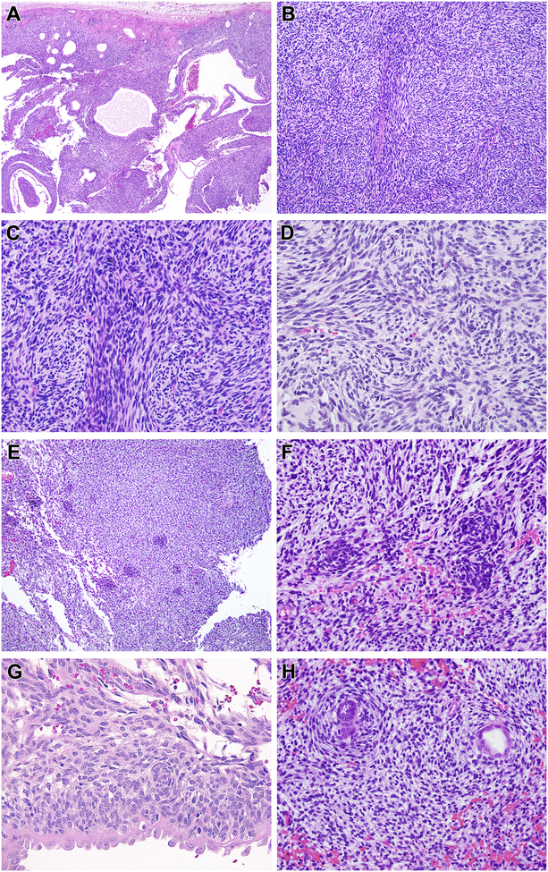
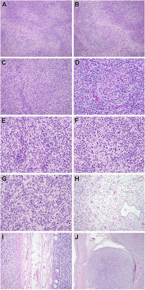
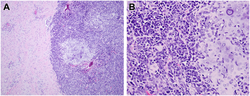
We describe 2 cases of a distinct sarcoma characterized by a novel MEIS1-NCOA2 gene fusion. This gene fusion was identified in the renal neoplasms of 2 adults (21-y-old male, 72-y-old female). Histologically, the resected renal neoplasms had a distinctively nodular appearance, and while one renal neoplasm was predominantly cystic, the other demonstrated solid architecture, invasion of perirenal fat, and renal sinus vasculature invasion. The neoplasms were characterized predominantly by monomorphic plump spindle cells arranged in vague fascicles with a whorling pattern; however, a more primitive small round cell component was also noted. Both neoplasms were mitotically active and one case showed necrosis. The neoplasms did not have a distinctive immunohistochemical profile, though both labeled for TLE1. The morphologic features are distinct from other sarcomas associated with NCOA2 gene fusions, including mesenchymal chondrosarcoma, congenital/infantile spindle cell rhabdomyosarcoma, and soft tissue angiofibroma. While we have minimal clinical follow-up, the aggressive histologic features of these neoplasms indicate malignant potential, thus warranting classification as a novel subtype of sarcoma.


Figures

Figure 1.
Figure 1.. Morphologic features of renal case 1 with MEIS1-NCOA2 fusion (index case).
This neoplasm had a dominant cystic appearance at low power (A), created by a solid spindle cell neoplasm associated with entrapped cystically-dilated renal tubules. The neoplasm had a nodular low power appearance (B) created by centrally located spindle cells with more abundant eosinophilic cytoplasm separated by primitive spindle to epithelioid cells with scant cytoplasm (C). The cytology in these areas overlapped with that of monophasic synovial sarcoma. The centrally located spindle cells demonstrated whorling (D). Small primitive cells arranged in clusters (E) and vague whorling similar to the spindle cells (F). The primitive neoplastic cells were cytologically distinct from the entrapped, cystically-dilated native renal tubular epithelium, which had more open, vesicular chromatin and hobnail appearance (G). In focal areas the spindle cells encircled native tubules in a concentric, “onion-skin” pattern (H).
Figure 2.
Figure 2.. Morphologic features of renal case 2 with MEIS1-NCOA2 fusion.
This solid cellular neoplasm had distinctive nodular variations in cellularity as evident at low power (A). In some areas, the nodularity was created by hypocellular fibrous zones separating more cellular nodules (B). The most consistent pattern, as seen in case 1, was that of centrally located spindle cells with eosinophilic cytoplasm forming whorls, surrounded by smaller cells with less cytoplasm (C, D); the smaller cells were packed in tight clusters, often centered on capillaries (E). Mitotic figures were readily identifiable in both components (F). In more cellular areas, the appearance resembles that of a small blue round cell tumor such as a primitive neuroectodermal tumor (PNET) with Homer Wright rosettes (G). This solid cellular neoplasm focally permeated between native renal tubules at its periphery but a significant cystic component was absent (H). The neoplasm invaded through the renal capsule to approach the adrenal gland (I) and showing invasion into the renal sinus (J).
Figure 3.
Figure 3.. MEIS1-NCOA2 gene fusion structure and molecular correlates.
Diagrammatic representation of MEIS1 gene on 2p14 fused to NCOA2 gene on 8q13.3 (A). RNAseq reads identified 2 fusion transcript isoforms, in which exon 7 of MEIS1 was fused (curved red arrow) to either exon 13 or 14 of NCOA2 (red box), likely due to alternative splicing. Representative sequences from RNAseq data demonstrating the fusion of exon 7 of MEIS1 to exon 13 of NCOA2, with emphasis on which protein domains are projected to be retained in the fusion oncoprotein (B). Unsupervised hierarchical clustering of RNAseq data showing that the MEIS1-NCOA2 renal sarcoma case 1 (blue) clustered together with the 2 mesenchymal chondrosarcomas with HEY1-NCOA2 fusion (green), separate from a soft tissue angiofibroma with AHRR-NCOA2 fusion (red)(C); gray lines refer to a large cohort of various sarcoma types studied on the same targeted RNAseq platform (C). Bar chart of HEY1 and AHRR mRNA expression showing upregulation in the mesenchymal chondrosarcomas and soft tissue angiofibroma, respectively, but not in MEIS1-NCOA2 positive renal sarcoma case 1 (D). Exon level expression of NCOA2 mRNA showing up-regulation of the 3’end of NCOA2 exons retained in the fusion transcript (breakpoints of renal sarcoma index case and 2 mesenchymal chondrosarcomas were identical in exons 13 and 14, green line; while breakpoint of angiofibroma in exon 16, red line)(E).
Figure 4.
Figure 4.. Break-apart FISH showing gene rearrangements in MEIS1 and NCOA2.
Renal sarcoma case 2 demonstrates rearrangement of MEIS1 (A) and NCOA2 (B) (red signal, centromeric; green signal, telomeric).
Figure 5.
Figure 5.. Histologic appearance of mesenchymal chondrosarcoma metastatic to the kidney for comparison.
At low power, one can appreciate the primitive small cells with associated nodules of cartilage to the right and entrapped kidney to the left (A). At high power (B), one can appreciate the cartilage and primitive small blue round cell appearance, which contrasts with the monomorphic spindle cell appearance of the MEIS1-NCOA2 renal sarcomas. This mesenchymal chondrosarcoma showed a NCOA2 gene rearrangement, but lacked evidence of MEIS1 gene abnormalities.

References

    1. Knezevich SR, Garnett MJ, Pysher TJ, et al. ETV6-NTRK3 gene fusions and trisomy 11 establish a histogenetic link between mesoblastic nephroma and congenital fibrosarcoma. Cancer Res 1998; 58:5046–5048. - PubMed
    1. Rubin BP, Chen CJ, Morgan TW, et al. Congenital mesoblastic nephroma t(12;15) is associated with ETV6-NTRK3 gene fusion: cytogenetic and molecular relationship to congenital (infantile) fibrosarcoma. Am J Pathol 1998; 153:1451–1458. - PMC - PubMed
    1. Argani P, Fritsch M, Kadkol SS, et al. Detection of the ETV6-NTRK3 chimeric RNA of infantile fibrosarcoma/cellular congenital mesoblastic nephroma in paraffin-embedded tissue: application to challenging pediatric renal stromal tumors. Mod Pathol 2000; 13:29–36. - PubMed
    1. Quezado M, Benjamin DR, Tsokos M EWS/FLI-1 fusion transcripts in three peripheral primitive neuroectodermal tumors of the kidney. Hum Pathol 1997; 28:767–771. - PubMed
    1. Jimenez RE, Folpe AL, Lapham RL, et al. Primary Ewing’s sarcoma/primitive neuroectodermal tumor of the kidney. A clinicopathologic and immunohistochemical analysis of 11 cases. Am J Surg Pathol 2002; 26:320–327. - PubMed

Publication types

MeSH terms