

Evaluation of Auramine O staining and conventional PCR for leprosy diagnosis: A comparative cross-sectional study from Ethiopia
- PMID: 30180155
- PMCID: PMC6138420
- DOI: 10.1371/journal.pntd.0006706
Evaluation of Auramine O staining and conventional PCR for leprosy diagnosis: A comparative cross-sectional study from Ethiopia
Abstract
Background: Diagnosis of leprosy mainly relies on clinical examination due to the inconsistent sensitivity and poor reproducibility of the current laboratory tests. Utilisation of alternative methods to the standard Ziehl Neelsen (ZN), Fite-Faraco (FF) and Haematoxylin and Eosin (H&E) staining procedures may eventually improve leprosy diagnosis.
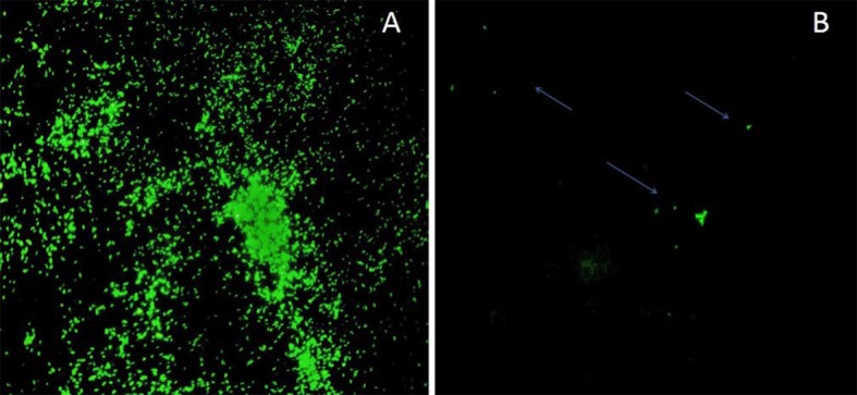
Methodology/principal findings: In this comparative study, the performance of the fluorescent Auramine O (AO) staining and polymerase chain reaction (PCR) was assessed with different skin samples using a combination of ZN, FF and H&E staining as the gold standard. AO, ZN, FF, H&E and PCR tests were performed on slit skin smears (SSS) and/or punch biopsies collected from 141 clinically confirmed leprosy cases and 28 non-leprosy skin samples. DNA was extracted from punch biopsies using two different methods with or without mechanical lysis. Sensitivities were 87.6%, 59.3% and 77% for H&E, ZN and FF, respectively, whereas it reached 65.5% and 77.9% for AO in SSS and tissue sections and 91.1% for PCR in tissue samples. Morover, samples with low bacillary index, sensitivity of AO staining (61.8%) was similar to FF (60%, p>0.05) and lower than PCR (86.6%, p<0.05). Sensitivity of PCR also increased (96.8%, p<0.05) when mechanical lysis was used during DNA extraction compared to enzymatic treatment alone (84.6%).
Conclusions/significance: Our results showed that for diagnostic purposes, analysis of skin section is more sensitive than SSS, especially for samples with low bacillary load. AO staining on SSS and tissue sections was not significantly better than other routine diagnostic tests but considerably more user friendly. The sensitivity of PCR was higher than current standard methods and increased when combined with more efficient DNA extraction using mechanical and chemical lysis. Therefore, we recommend AO staining for the diagnosis of leprosy in lower health facilities such as health centres and district hospitals and PCR diagnosis at referral level and research centres.
Conflict of interest statement
The authors have declared that no competing interest exist.
Figures

References
-
- Britton WJ, Lockwood DN. Leprosy. Lancet (London, England). 2004; 363: 1209–19. - PubMed
-
- Ridley DS, Jopling WH. A classification of leprosy for research purposes. Leprosy review. 1962; 33: 119–28. - PubMed
-
- FMoH. Tuberculosis, Leprosy andTB/HIV Prevention and Control Programme Addis Ababa: Federal Ministry of Health Ethiopia; 2008.
-
- WHO. WHO model prescribing information: drugs used in leprosy Geneva: World Health Organization; 1998.
-
- WHO. Global leprosy update, 2016: accelerating reduction of disease burden Geneva: World Health Organization; 2017. - PubMed
Publication types
MeSH terms
Substances
LinkOut - more resources
Full Text Sources
Other Literature Sources
Medical
